6QRZ

Crystal structure of R2-like ligand-binding oxidase from Sulfolobus acidocaldarius solved by 3D micro-crystal electron diffraction


Experimental Data Snapshot

  • Method: ELECTRON CRYSTALLOGRAPHY
  • Resolution: 3.00 Å
  • R-Value Free: 0.335 
  • R-Value Work: 0.318 
  • R-Value Observed: 0.319 

wwPDB Validation   3D Report Full Report


This is version 1.2 of the entry. See complete history


Literature

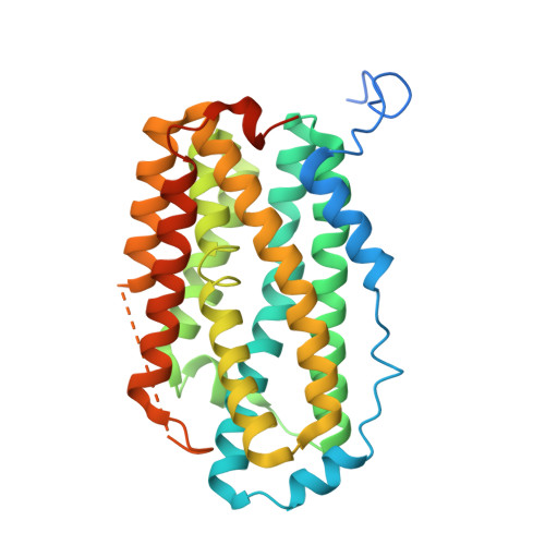
Solving a new R2lox protein structure by microcrystal electron diffraction.

Xu, H.Lebrette, H.Clabbers, M.T.B.Zhao, J.Griese, J.J.Zou, X.Hogbom, M.

(2019) Sci Adv 5: eaax4621-eaax4621

  • DOI: https://doi.org/10.1126/sciadv.aax4621
  • Primary Citation of Related Structures:  
    6QRZ

  • PubMed Abstract: 

    Microcrystal electron diffraction (MicroED) has recently shown potential for structural biology. It enables the study of biomolecules from micrometer-sized 3D crystals that are too small to be studied by conventional x-ray crystallography. However, to date, MicroED has only been applied to redetermine protein structures that had already been solved previously by x-ray diffraction. Here, we present the first new protein structure-an R2lox enzyme-solved using MicroED. The structure was phased by molecular replacement using a search model of 35% sequence identity. The resulting electrostatic scattering potential map at 3.0-Å resolution was of sufficient quality to allow accurate model building and refinement. The dinuclear metal cofactor could be located in the map and was modeled as a heterodinuclear Mn/Fe center based on previous studies. Our results demonstrate that MicroED has the potential to become a widely applicable tool for revealing novel insights into protein structure and function.


  • Organizational Affiliation

    Department of Materials and Environmental Chemistry, Stockholm University, 10691 Stockholm, Sweden.


Macromolecules
Find similar proteins by:  (by identity cutoff)  |  3D Structure
Entity ID: 1
MoleculeChains Sequence LengthOrganismDetailsImage
Ribonucleoside-diphosphate reductase328Sulfolobus acidocaldarius DSM 639Mutation(s): 0 
Gene Names: Saci_2188
EC: 1.17.4.1
UniProt
Find proteins for Q4J6V7 (Sulfolobus acidocaldarius (strain ATCC 33909 / DSM 639 / JCM 8929 / NBRC 15157 / NCIMB 11770))
Explore Q4J6V7 
Go to UniProtKB:  Q4J6V7
Entity Groups  
Sequence Clusters30% Identity50% Identity70% Identity90% Identity95% Identity100% Identity
UniProt GroupQ4J6V7
Sequence Annotations
Expand
  • Reference Sequence
Small Molecules
Ligands 2 Unique
IDChains Name / Formula / InChI Key2D Diagram3D Interactions
FE
Query on FE

Download Ideal Coordinates CCD File 
C [auth A]FE (III) ION
Fe
VTLYFUHAOXGGBS-UHFFFAOYSA-N
MN3
Query on MN3

Download Ideal Coordinates CCD File 
B [auth A]MANGANESE (III) ION
Mn
MMIPFLVOWGHZQD-UHFFFAOYSA-N
Experimental Data & Validation

Experimental Data

  • Method: ELECTRON CRYSTALLOGRAPHY
  • Resolution: 3.00 Å
  • R-Value Free: 0.335 
  • R-Value Work: 0.318 
  • R-Value Observed: 0.319 
  • Space Group: P 21 21 2
Unit Cell:
Length ( Å )Angle ( ˚ )
a = 63.31α = 90
b = 108.93β = 90
c = 48.17γ = 90
Software Package:
Software NamePurpose
PHENIXrefinement
XDSdata reduction
XSCALEdata scaling
PHASERphasing
Cootmodel building
EM Software:
TaskSoftware PackageVersion
MODEL REFINEMENTPHENIX

Structure Validation

View Full Validation Report


Entry History & Funding Information

Deposition Data


Funding OrganizationLocationGrant Number
Swedish Research CouncilSweden2017-04018
Knut and Alice Wallenberg FoundationSwedenWallenberg Academy Fellows 2017.0275
Knut and Alice Wallenberg FoundationSweden3DEM-NATUR 2012.0112
Swedish Research CouncilSwedenVR Starting Grant 2017-05333

Revision History  (Full details and data files)

  • Version 1.0: 2019-04-24
    Type: Initial release
  • Version 1.1: 2019-09-04
    Changes: Data collection, Database references
  • Version 1.2: 2019-09-11
    Changes: Data collection, Database references