6QRJ

Crystal structure of ShkA full-length in complex with AMPPNP


Experimental Data Snapshot

  • Method: X-RAY DIFFRACTION
  • Resolution: 2.65 Å
  • R-Value Free: 0.275 
  • R-Value Work: 0.217 
  • R-Value Observed: 0.220 

wwPDB Validation   3D Report Full Report


Ligand Structure Quality Assessment 


This is version 1.2 of the entry. See complete history


Literature

Hybrid histidine kinase activation by cyclic di-GMP-mediated domain liberation.

Dubey, B.N.Agustoni, E.Bohm, R.Kaczmarczyk, A.Mangia, F.von Arx, C.Jenal, U.Hiller, S.Plaza-Menacho, I.Schirmer, T.

(2020) Proc Natl Acad Sci U S A 117: 1000-1008

  • DOI: https://doi.org/10.1073/pnas.1911427117
  • Primary Citation of Related Structures:  
    6QRJ, 6QRL

  • PubMed Abstract: 

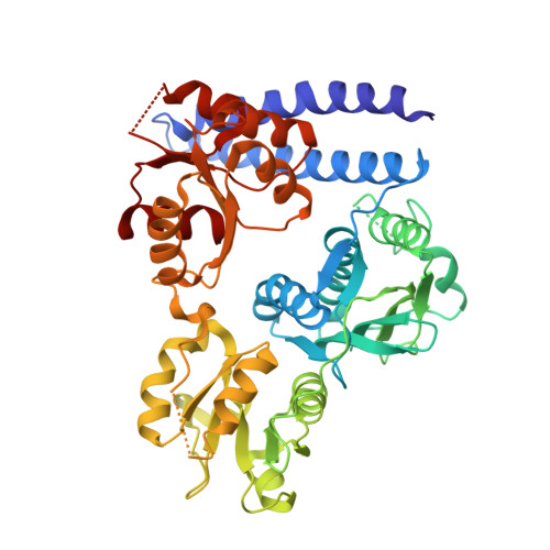
    Cytosolic hybrid histidine kinases (HHKs) constitute major signaling nodes that control various biological processes, but their input signals and how these are processed are largely unknown. In Caulobacter crescentus , the HHK ShkA is essential for accurate timing of the G1-S cell cycle transition and is regulated by the corresponding increase in the level of the second messenger c-di-GMP. Here, we use a combination of X-ray crystallography, NMR spectroscopy, functional analyses, and kinetic modeling to reveal the regulatory mechanism of ShkA. In the absence of c-di-GMP, ShkA predominantly adopts a compact domain arrangement that is catalytically inactive. C-di-GMP binds to the dedicated pseudoreceiver domain Rec1, thereby liberating the canonical Rec2 domain from its central position where it obstructs the large-scale motions required for catalysis. Thus, c-di-GMP cannot only stabilize domain interactions, but also engage in domain dissociation to allosterically invoke a downstream effect. Enzyme kinetics data are consistent with conformational selection of the ensemble of active domain constellations by the ligand and show that autophosphorylation is a reversible process.


  • Organizational Affiliation

    Structural Biology, Biozentrum, University of Basel, 4056 Basel, Switzerland.


Macromolecules
Find similar proteins by:  (by identity cutoff)  |  3D Structure
Entity ID: 1
MoleculeChains Sequence LengthOrganismDetailsImage
Hybrid kinase484Caulobacter vibrioides CB15Mutation(s): 0 
Gene Names: shkACC_0138CC0138
UniProt
Find proteins for Q9ABT2 (Caulobacter vibrioides (strain ATCC 19089 / CB15))
Explore Q9ABT2 
Go to UniProtKB:  Q9ABT2
Entity Groups  
Sequence Clusters30% Identity50% Identity70% Identity90% Identity95% Identity100% Identity
UniProt GroupQ9ABT2
Sequence Annotations
Expand
  • Reference Sequence
Small Molecules
Experimental Data & Validation

Experimental Data

  • Method: X-RAY DIFFRACTION
  • Resolution: 2.65 Å
  • R-Value Free: 0.275 
  • R-Value Work: 0.217 
  • R-Value Observed: 0.220 
  • Space Group: P 43 2 2
Unit Cell:
Length ( Å )Angle ( ˚ )
a = 64.504α = 90
b = 64.504β = 90
c = 260.209γ = 90
Software Package:
Software NamePurpose
REFMACrefinement
XDSdata reduction
XSCALEdata scaling
PDB_EXTRACTdata extraction
PHASERphasing

Structure Validation

View Full Validation Report


Ligand Structure Quality Assessment 


Entry History & Funding Information

Deposition Data


Funding OrganizationLocationGrant Number
Swiss National Science FoundationSwitzerland31003A_166652

Revision History  (Full details and data files)

  • Version 1.0: 2020-01-08
    Type: Initial release
  • Version 1.1: 2020-01-22
    Changes: Database references
  • Version 1.2: 2024-01-24
    Changes: Data collection, Database references, Refinement description