6QPL

Crystal structure of Spindlin1 in complex with the inhibitor MS31


Experimental Data Snapshot

  • Method: X-RAY DIFFRACTION
  • Resolution: 1.60 Å
  • R-Value Free: 0.208 
  • R-Value Work: 0.190 
  • R-Value Observed: 0.191 

wwPDB Validation   3D Report Full Report


Ligand Structure Quality Assessment 


This is version 1.2 of the entry. See complete history


Literature

Discovery of a Potent and Selective Fragment-like Inhibitor of Methyllysine Reader Protein Spindlin 1 (SPIN1).

Xiong, Y.Greschik, H.Johansson, C.Seifert, L.Bacher, J.Park, K.S.Babault, N.Martini, M.Fagan, V.Li, F.Chau, I.Christott, T.Dilworth, D.Barsyte-Lovejoy, D.Vedadi, M.Arrowsmith, C.H.Brennan, P.Fedorov, O.Jung, M.Farnie, G.Liu, J.Oppermann, U.Schule, R.Jin, J.

(2019) J Med Chem 62: 8996-9007

  • DOI: https://doi.org/10.1021/acs.jmedchem.9b00522
  • Primary Citation of Related Structures:  
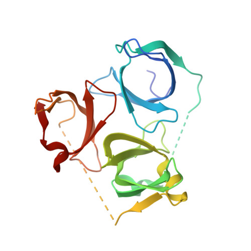
    6QPL

  • PubMed Abstract: 

    By screening an epigenetic compound library, we identified that UNC0638, a highly potent inhibitor of the histone methyltransferases G9a and GLP, was a weak inhibitor of SPIN1 (spindlin 1), a methyllysine reader protein. Our optimization of this weak hit resulted in the discovery of a potent, selective, and cell-active SPIN1 inhibitor, compound 3 (MS31). Compound 3 potently inhibited binding of trimethyllysine-containing peptides to SPIN1, displayed high binding affinity, was highly selective for SPIN1 over other epigenetic readers and writers, directly engaged SPIN1 in cells, and was not toxic to nontumorigenic cells. The crystal structure of the SPIN1-compound 3 complex indicated that it selectively binds tudor domain II of SPIN1. We also designed a structurally similar but inactive compound 4 (MS31N) as a negative control. Our results have demonstrated for the first time that potent, selective, and cell-active fragment-like inhibitors can be generated by targeting a single tudor domain.


  • Organizational Affiliation

    Mount Sinai Center for Therapeutics Discovery, Departments of Pharmacological Sciences and Oncological Sciences, Tisch Cancer Institute , Icahn School of Medicine at Mount Sinai , New York , New York 10029 , United States.


Macromolecules
Find similar proteins by:  (by identity cutoff)  |  3D Structure
Entity ID: 1
MoleculeChains Sequence LengthOrganismDetailsImage
Spindlin-1A [auth B]222Homo sapiensMutation(s): 0 
Gene Names: SPIN1OCRSPIN
UniProt & NIH Common Fund Data Resources
Find proteins for Q9Y657 (Homo sapiens)
Explore Q9Y657 
Go to UniProtKB:  Q9Y657
PHAROS:  Q9Y657
GTEx:  ENSG00000106723 
Entity Groups  
Sequence Clusters30% Identity50% Identity70% Identity90% Identity95% Identity100% Identity
UniProt GroupQ9Y657
Sequence Annotations
Expand
  • Reference Sequence
Small Molecules
Ligands 6 Unique
IDChains Name / Formula / InChI Key2D Diagram3D Interactions
JC5
Query on JC5

Download Ideal Coordinates CCD File 
F [auth B][3-(aminomethyl)-5-[3-(1,3-dihydroisoindol-2-yl)propoxy]-4-methoxy-phenyl]methanamine
C20 H27 N3 O2
XQQZNYFJATXWON-UHFFFAOYSA-N
MPD
Query on MPD

Download Ideal Coordinates CCD File 
B,
C [auth B]
(4S)-2-METHYL-2,4-PENTANEDIOL
C6 H14 O2
SVTBMSDMJJWYQN-YFKPBYRVSA-N
PO4
Query on PO4

Download Ideal Coordinates CCD File 
H [auth B]PHOSPHATE ION
O4 P
NBIIXXVUZAFLBC-UHFFFAOYSA-K
GOL
Query on GOL

Download Ideal Coordinates CCD File 
I [auth B]GLYCEROL
C3 H8 O3
PEDCQBHIVMGVHV-UHFFFAOYSA-N
DMS
Query on DMS

Download Ideal Coordinates CCD File 
D [auth B],
E [auth B]
DIMETHYL SULFOXIDE
C2 H6 O S
IAZDPXIOMUYVGZ-UHFFFAOYSA-N
GLY
Query on GLY

Download Ideal Coordinates CCD File 
G [auth B]GLYCINE
C2 H5 N O2
DHMQDGOQFOQNFH-UHFFFAOYSA-N
Binding Affinity Annotations 
IDSourceBinding Affinity
JC5 Binding MOAD:  6QPL IC50: 77 (nM) from 1 assay(s)
Experimental Data & Validation

Experimental Data

  • Method: X-RAY DIFFRACTION
  • Resolution: 1.60 Å
  • R-Value Free: 0.208 
  • R-Value Work: 0.190 
  • R-Value Observed: 0.191 
  • Space Group: P 43 21 2
Unit Cell:
Length ( Å )Angle ( ˚ )
a = 115.3α = 90
b = 115.3β = 90
c = 43.59γ = 90
Software Package:
Software NamePurpose
XDSdata reduction
Aimlessdata scaling
PHASERphasing
PHENIXrefinement
PDB_EXTRACTdata extraction

Structure Validation

View Full Validation Report


Ligand Structure Quality Assessment 


Entry History & Funding Information

Deposition Data


Funding OrganizationLocationGrant Number
United Kingdom--

Revision History  (Full details and data files)

  • Version 1.0: 2019-07-17
    Type: Initial release
  • Version 1.1: 2019-11-06
    Changes: Data collection, Database references
  • Version 1.2: 2024-01-24
    Changes: Data collection, Database references, Refinement description