6QE2

Crystal structure of Paleococcus ferrophilus monoacylglycerol lipase.


Experimental Data Snapshot

  • Method: X-RAY DIFFRACTION
  • Resolution: 1.75 Å
  • R-Value Free: 0.198 
  • R-Value Work: 0.171 
  • R-Value Observed: 0.172 

wwPDB Validation   3D Report Full Report


Ligand Structure Quality Assessment 


This is version 1.2 of the entry. See complete history


Literature

Structure and Dynamics of an Archeal Monoglyceride Lipase from Palaeococcus ferrophilus as Revealed by Crystallography and In Silico Analysis.

Labar, G.Brandt, N.Flaba, A.Wouters, J.Leherte, L.

(2021) Biomolecules 11

  • DOI: https://doi.org/10.3390/biom11040533
  • Primary Citation of Related Structures:  
    6QE2

  • PubMed Abstract: 

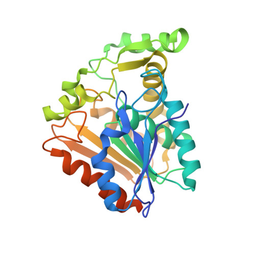
    The crystallographic analysis of a lipase from Palaeococcus ferrophilus (PFL) previously annotated as a lysophospholipase revealed high structural conservation with other monoglyceride lipases, in particular in the lid domain and substrate binding pockets. In agreement with this observation, PFL was shown to be active on various monoacylglycerols. Molecular Dynamics (MD) studies performed in the absence and in the presence of ligands further allowed characterization of the dynamics of this system and led to a systematic closure of the lid compared to the crystal structure. However, the presence of ligands in the acyl-binding pocket stabilizes intermediate conformations compared to the crystal and totally closed structures. Several lid-stabilizing or closure elements were highlighted, i.e., hydrogen bonds between Ser117 and Ile204 or Asn142 and its facing amino acid lid residues, as well as Phe123. Thus, based on this complementary crystallographic and MD approach, we suggest that the crystal structure reported herein represents an open conformation, at least partially, of the PFL, which is likely stabilized by the ligand, and it brings to light several key structural features prone to participate in the closure of the lid.


  • Organizational Affiliation

    Institut de Recherches Labiris, 1 Avenue E. Gryson, B-1070 Bruxelles, Belgium.


Macromolecules
Find similar proteins by:  (by identity cutoff)  |  3D Structure
Entity ID: 1
MoleculeChains Sequence LengthOrganismDetailsImage
Monoacylglycerol lipase
A, B
283Palaeococcus ferrophilusMutation(s): 0 
EC: 3.1.1.23
Entity Groups  
Sequence Clusters30% Identity50% Identity70% Identity90% Identity95% Identity100% Identity
Sequence Annotations
Expand
  • Reference Sequence
Experimental Data & Validation

Experimental Data

  • Method: X-RAY DIFFRACTION
  • Resolution: 1.75 Å
  • R-Value Free: 0.198 
  • R-Value Work: 0.171 
  • R-Value Observed: 0.172 
  • Space Group: C 1 2 1
Unit Cell:
Length ( Å )Angle ( ˚ )
a = 120.6α = 90
b = 75.46β = 128.29
c = 85.44γ = 90
Software Package:
Software NamePurpose
PHENIXrefinement
XDSdata reduction
XSCALEdata scaling
PHASERphasing
PDB_EXTRACTdata extraction

Structure Validation

View Full Validation Report


Ligand Structure Quality Assessment 


Entry History & Funding Information

Deposition Data


Funding OrganizationLocationGrant Number
BelgiumBAG-Belgium 20151139
BelgiumBAG-Belgium 20161175

Revision History  (Full details and data files)

  • Version 1.0: 2020-02-05
    Type: Initial release
  • Version 1.1: 2021-05-19
    Changes: Database references
  • Version 1.2: 2024-01-24
    Changes: Data collection, Database references, Refinement description