6QAZ

Crystal structure of gp41-1 intein


Experimental Data Snapshot

  • Method: X-RAY DIFFRACTION
  • Resolution: 1.02 Å
  • R-Value Free: 0.150 
  • R-Value Work: 0.121 
  • R-Value Observed: 0.123 

wwPDB Validation   3D Report Full Report


This is version 1.3 of the entry. See complete history


Literature

The crystal structure of the naturally split gp41-1 intein guides the engineering of orthogonal split inteins from cis-splicing inteins.

Beyer, H.M.Mikula, K.M.Li, M.Wlodawer, A.Iwai, H.

(2020) FEBS J 287: 1886-1898

  • DOI: https://doi.org/10.1111/febs.15113
  • Primary Citation of Related Structures:  
    6QAZ

  • PubMed Abstract: 

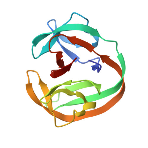
    Protein trans-splicing catalyzed by split inteins has increasingly become useful as a protein engineering tool. We solved the 1.0 Å-resolution crystal structure of a fused variant from the naturally split gp41-1 intein, previously identified from environmental metagenomic sequence data. The structure of the 125-residue gp41-1 intein revealed a compact pseudo-C2-symmetry commonly found in the Hedgehog/Intein superfamily with extensive charge-charge interactions between the split N- and C-terminal intein fragments that are common among naturally occurring split inteins. We successfully created orthogonal split inteins by engineering a similar charge network into the same region of a cis-splicing intein. This strategy could be applicable for creating novel natural-like split inteins from other, more prevalent cis-splicing inteins. DATABASE: Structural data are available in the RCSB Protein Data Bank under the accession number 6QAZ.


  • Organizational Affiliation

    Research Program in Structural Biology and Biophysics, Helsinki Life Science Institute-Institute of Biotechnology, University of Helsinki, Finland.


Macromolecules
Find similar proteins by:  (by identity cutoff)  |  3D Structure
Entity ID: 1
MoleculeChains Sequence LengthOrganismDetailsImage
cyanophage-like gp41-1 intein128metagenomeMutation(s): 0 
Entity Groups  
Sequence Clusters30% Identity50% Identity70% Identity90% Identity95% Identity100% Identity
Sequence Annotations
Expand
  • Reference Sequence
Experimental Data & Validation

Experimental Data

  • Method: X-RAY DIFFRACTION
  • Resolution: 1.02 Å
  • R-Value Free: 0.150 
  • R-Value Work: 0.121 
  • R-Value Observed: 0.123 
  • Space Group: I 2 2 2
Unit Cell:
Length ( Å )Angle ( ˚ )
a = 48.809α = 90
b = 69.986β = 90
c = 71.236γ = 90
Software Package:
Software NamePurpose
REFMACrefinement
HKL-3000data reduction
HKL-3000data scaling
PHASERphasing

Structure Validation

View Full Validation Report


Entry History & Funding Information

Deposition Data


Funding OrganizationLocationGrant Number
Academy of FinlandFinland137995, 277335
Novo Nordisk FoundationFinlandNNF17OC0025402
Sigrid Juselius FoundationFinland--
National Institutes of Health/National Cancer Institute (NIH/NCI)United StatesHHSN261200800001E

Revision History  (Full details and data files)

  • Version 1.0: 2019-11-13
    Type: Initial release
  • Version 1.1: 2020-05-13
    Changes: Database references
  • Version 1.2: 2022-03-30
    Changes: Author supporting evidence, Database references
  • Version 1.3: 2024-01-24
    Changes: Data collection, Refinement description