6Q9M

Central Fibronectin-III array of RIM-binding protein


Experimental Data Snapshot

  • Method: X-RAY DIFFRACTION
  • Resolution: 2.45 Å
  • R-Value Free: 0.260 
  • R-Value Work: 0.211 
  • R-Value Observed: 0.213 

wwPDB Validation   3D Report Full Report


This is version 1.2 of the entry. See complete history


Literature

RIM-binding protein couples synaptic vesicle recruitment to release sites.

Petzoldt, A.G.Gotz, T.W.B.Driller, J.H.Lutzkendorf, J.Reddy-Alla, S.Matkovic-Rachid, T.Liu, S.Knoche, E.Mertel, S.Ugorets, V.Lehmann, M.Ramesh, N.Beuschel, C.B.Kuropka, B.Freund, C.Stelzl, U.Loll, B.Liu, F.Wahl, M.C.Sigrist, S.J.

(2020) J Cell Biol 219

  • DOI: https://doi.org/10.1083/jcb.201902059
  • Primary Citation of Related Structures:  
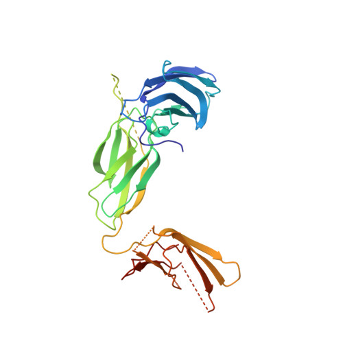
    6Q9M

  • PubMed Abstract: 

    At presynaptic active zones, arrays of large conserved scaffold proteins mediate fast and temporally precise release of synaptic vesicles (SVs). SV release sites could be identified by clusters of Munc13, which allow SVs to dock in defined nanoscale relation to Ca2+ channels. We here show in Drosophila that RIM-binding protein (RIM-BP) connects release sites physically and functionally to the ELKS family Bruchpilot (BRP)-based scaffold engaged in SV recruitment. The RIM-BP N-terminal domain, while dispensable for SV release site organization, was crucial for proper nanoscale patterning of the BRP scaffold and needed for SV recruitment of SVs under strong stimulation. Structural analysis further showed that the RIM-BP fibronectin domains form a "hinge" in the protein center, while the C-terminal SH3 domain tandem binds RIM, Munc13, and Ca2+ channels release machinery collectively. RIM-BPs' conserved domain architecture seemingly provides a relay to guide SVs from membrane far scaffolds into membrane close release sites.


  • Organizational Affiliation

    Freie Universität Berlin, Institute for Biology and Genetics, Berlin, Germany.


Macromolecules
Find similar proteins by:  (by identity cutoff)  |  3D Structure
Entity ID: 1
MoleculeChains Sequence LengthOrganismDetailsImage
RIM-binding protein, isoform F
A, B
303Drosophila melanogasterMutation(s): 0 
Gene Names: RbpCG14867CG31302CG5143CG5152Dmel\CG43073DRBPRBPCG43073Dmel_CG43073
UniProt
Find proteins for A0A0B4JDC9 (Drosophila melanogaster)
Explore A0A0B4JDC9 
Go to UniProtKB:  A0A0B4JDC9
Entity Groups  
Sequence Clusters30% Identity50% Identity70% Identity90% Identity95% Identity100% Identity
UniProt GroupA0A0B4JDC9
Sequence Annotations
Expand
  • Reference Sequence
Small Molecules
Ligands 1 Unique
IDChains Name / Formula / InChI Key2D Diagram3D Interactions
PO4
Query on PO4

Download Ideal Coordinates CCD File 
C [auth A]PHOSPHATE ION
O4 P
NBIIXXVUZAFLBC-UHFFFAOYSA-K
Experimental Data & Validation

Experimental Data

  • Method: X-RAY DIFFRACTION
  • Resolution: 2.45 Å
  • R-Value Free: 0.260 
  • R-Value Work: 0.211 
  • R-Value Observed: 0.213 
  • Space Group: P 1 21 1
Unit Cell:
Length ( Å )Angle ( ˚ )
a = 51.095α = 90
b = 80.547β = 99.03
c = 93.077γ = 90
Software Package:
Software NamePurpose
PHENIXrefinement
XDSdata reduction
XSCALEdata scaling
SHARPphasing
BUCCANEERmodel building

Structure Validation

View Full Validation Report


Entry History & Funding Information

Deposition Data


Funding OrganizationLocationGrant Number
German Research FoundationGermanySFB958

Revision History  (Full details and data files)

  • Version 1.0: 2020-01-15
    Type: Initial release
  • Version 1.1: 2020-04-15
    Changes: Database references
  • Version 1.2: 2020-05-13
    Changes: Database references