6Q54

Structure of GluA2 ligand-binding domain (S1S2J) in complex with the agonist (S)-2-Amino-3-(1-ethyl-4-hydroxy-1H-1,2,3-triazol-5-yl)propanoic acid at 1.4 A resolution


Experimental Data Snapshot

  • Method: X-RAY DIFFRACTION
  • Resolution: 1.40 Å
  • R-Value Free: 0.163 
  • R-Value Work: 0.144 
  • R-Value Observed: 0.145 

wwPDB Validation   3D Report Full Report


Ligand Structure Quality Assessment 


This is version 1.2 of the entry. See complete history


Literature

Use of the 4-Hydroxytriazole Moiety as a Bioisosteric Tool in the Development of Ionotropic Glutamate Receptor Ligands.

Sainas, S.Temperini, P.Farnsworth, J.C.Yi, F.Mollerud, S.Jensen, A.A.Nielsen, B.Passoni, A.Kastrup, J.S.Hansen, K.B.Boschi, D.Pickering, D.S.Clausen, R.P.Lolli, M.L.

(2019) J Med Chem 62: 4467-4482

  • DOI: https://doi.org/10.1021/acs.jmedchem.8b01986
  • Primary Citation of Related Structures:  
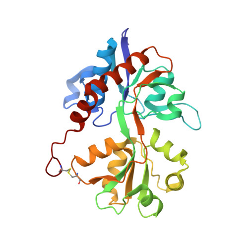
    6Q54, 6Q60

  • PubMed Abstract: 

    We report a series of glutamate and aspartate analogues designed using the hydroxy-1,2,3-triazole moiety as a bioisostere for the distal carboxylic acid. Compound 6b showed unprecedented selectivity among ( S)-2-amino-3-(3-hydroxy-5-methyl-4-isoxazolyl)propionic acid (AMPA) receptor subtypes, confirmed also by an unusual binding mode observed for the crystal structures in complex with the AMPA receptor GluA2 agonist-binding domain. Here, a methionine (Met729) was highly disordered compared to previous agonist-bound structures. This observation provides a possible explanation for the pharmacological profile. In the structure with 7a, an unusual organization of water molecules around the bioisostere arises compared to previous structures of ligands with other bioisosteres. Aspartate analogue 8 with the hydroxy-1,2,3-triazole moiety directly attached to glycine was unexpectedly able to activate both the glutamate and glycine agonist-binding sites of the N-methyl-d-aspartic acid receptor. These observations demonstrate novel features that arise when employing a hydroxytriazole moiety as a bioisostere for the distal carboxylic acid in glutamate receptor agonists.


  • Organizational Affiliation

    Department of Drug Science and Technology , University of Turin , via P.Giuria 9 , 10125 Turin , Italy.


Macromolecules
Find similar proteins by:  (by identity cutoff)  |  3D Structure
Entity ID: 1
MoleculeChains Sequence LengthOrganismDetailsImage
Glutamate receptor 2
A, B
264Rattus norvegicusMutation(s): 0 
Gene Names: Gria2Glur2
UniProt
Find proteins for P19491 (Rattus norvegicus)
Explore P19491 
Go to UniProtKB:  P19491
Entity Groups  
Sequence Clusters30% Identity50% Identity70% Identity90% Identity95% Identity100% Identity
UniProt GroupP19491
Sequence Annotations
Expand
  • Reference Sequence
Small Molecules
Ligands 8 Unique
IDChains Name / Formula / InChI Key2D Diagram3D Interactions
HJ8
Query on HJ8

Download Ideal Coordinates CCD File 
I [auth A],
V [auth B]
(2~{S})-2-azanyl-3-(3-ethyl-5-oxidanyl-1,2,3-triazol-4-yl)propanoic acid
C7 H12 N4 O3
ROBLGUMDXHEHOO-BYPYZUCNSA-N
CIT
Query on CIT

Download Ideal Coordinates CCD File 
O [auth A]CITRIC ACID
C6 H8 O7
KRKNYBCHXYNGOX-UHFFFAOYSA-N
PGE
Query on PGE

Download Ideal Coordinates CCD File 
K [auth A],
Z [auth B]
TRIETHYLENE GLYCOL
C6 H14 O4
ZIBGPFATKBEMQZ-UHFFFAOYSA-N
PEG
Query on PEG

Download Ideal Coordinates CCD File 
BA [auth B]DI(HYDROXYETHYL)ETHER
C4 H10 O3
MTHSVFCYNBDYFN-UHFFFAOYSA-N
SO4
Query on SO4

Download Ideal Coordinates CCD File 
C [auth A]
D [auth A]
E [auth A]
F [auth A]
G [auth A]
C [auth A],
D [auth A],
E [auth A],
F [auth A],
G [auth A],
H [auth A],
R [auth B],
S [auth B],
T [auth B],
U [auth B]
SULFATE ION
O4 S
QAOWNCQODCNURD-UHFFFAOYSA-L
GOL
Query on GOL

Download Ideal Coordinates CCD File 
J [auth A],
W [auth B],
X [auth B],
Y [auth B]
GLYCEROL
C3 H8 O3
PEDCQBHIVMGVHV-UHFFFAOYSA-N
CL
Query on CL

Download Ideal Coordinates CCD File 
L [auth A],
M [auth A],
N [auth A]
CHLORIDE ION
Cl
VEXZGXHMUGYJMC-UHFFFAOYSA-M
LI
Query on LI

Download Ideal Coordinates CCD File 
AA [auth B],
P [auth A],
Q [auth A]
LITHIUM ION
Li
HBBGRARXTFLTSG-UHFFFAOYSA-N
Experimental Data & Validation

Experimental Data

  • Method: X-RAY DIFFRACTION
  • Resolution: 1.40 Å
  • R-Value Free: 0.163 
  • R-Value Work: 0.144 
  • R-Value Observed: 0.145 
  • Space Group: P 21 21 2
Unit Cell:
Length ( Å )Angle ( ˚ )
a = 98.331α = 90
b = 121.738β = 90
c = 47.134γ = 90
Software Package:
Software NamePurpose
PHENIXrefinement
SCALAdata scaling
PDB_EXTRACTdata extraction
XDSdata reduction
PHASERphasing

Structure Validation

View Full Validation Report


Ligand Structure Quality Assessment 


Entry History 

Deposition Data

Revision History  (Full details and data files)

  • Version 1.0: 2019-04-17
    Type: Initial release
  • Version 1.1: 2019-05-22
    Changes: Data collection, Database references
  • Version 1.2: 2024-01-24
    Changes: Data collection, Database references, Derived calculations, Refinement description