6PXU

Crystal structure of human GalNAc-T12 bound to a diglycosylated peptide, Mn2+, and UDP


Experimental Data Snapshot

  • Method: X-RAY DIFFRACTION
  • Resolution: 2.01 Å
  • R-Value Free: 0.219 
  • R-Value Work: 0.171 
  • R-Value Observed: 0.173 

wwPDB Validation   3D Report Full Report

Currently 6PXU does not have a validation slider image.


This is version 2.1 of the entry. See complete history


Literature

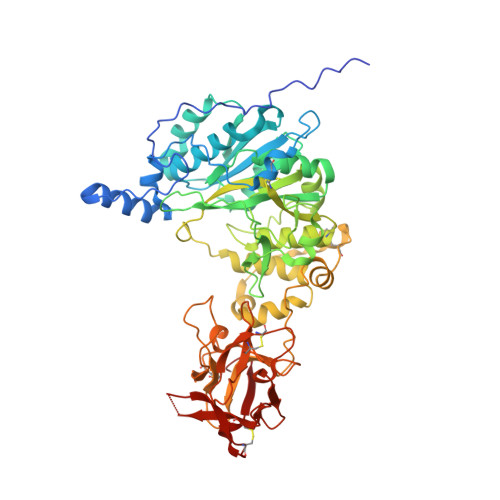
The structure of the colorectal cancer-associated enzyme GalNAc-T12 reveals how nonconserved residues dictate its function.

Fernandez, A.J.Daniel, E.J.P.Mahajan, S.P.Gray, J.J.Gerken, T.A.Tabak, L.A.Samara, N.L.

(2019) Proc Natl Acad Sci U S A 116: 20404-20410

  • DOI: https://doi.org/10.1073/pnas.1902211116
  • Primary Citation of Related Structures:  
    6PXU

  • PubMed Abstract: 

    Polypeptide N- acetylgalactosaminyl transferases (GalNAc-Ts) initiate mucin type O -glycosylation by catalyzing the transfer of N -acetylgalactosamine (GalNAc) to Ser or Thr on a protein substrate. Inactive and partially active variants of the isoenzyme GalNAc-T12 are present in subsets of patients with colorectal cancer, and several of these variants alter nonconserved residues with unknown functions. While previous biochemical studies have demonstrated that GalNAc-T12 selects for peptide and glycopeptide substrates through unique interactions with its catalytic and lectin domains, the molecular basis for this distinct substrate selectivity remains elusive. Here we examine the molecular basis of the activity and substrate selectivity of GalNAc-T12. The X-ray crystal structure of GalNAc-T12 in complex with a di-glycosylated peptide substrate reveals how a nonconserved GalNAc binding pocket in the GalNAc-T12 catalytic domain dictates its unique substrate selectivity. In addition, the structure provides insight into how colorectal cancer mutations disrupt the activity of GalNAc-T12 and illustrates how the rules dictating GalNAc-T12 function are distinct from those for other GalNAc-Ts.


  • Organizational Affiliation

    Section on Biological Chemistry, National Institute of Dental and Craniofacial Research, National Institutes of Health, Bethesda, MD 20892.


Macromolecules
Find similar proteins by:  (by identity cutoff)  |  3D Structure
Entity ID: 1
MoleculeChains Sequence LengthOrganismDetailsImage
Polypeptide N-acetylgalactosaminyltransferase 12
A, B
543Homo sapiensMutation(s): 0 
Gene Names: GALNT12
EC: 2.4.1.41
UniProt & NIH Common Fund Data Resources
Find proteins for Q8IXK2 (Homo sapiens)
Explore Q8IXK2 
Go to UniProtKB:  Q8IXK2
PHAROS:  Q8IXK2
GTEx:  ENSG00000119514 
Entity Groups  
Sequence Clusters30% Identity50% Identity70% Identity90% Identity95% Identity100% Identity
UniProt GroupQ8IXK2
Sequence Annotations
Expand
  • Reference Sequence

Find similar proteins by:  Sequence   |   3D Structure  

Entity ID: 2
MoleculeChains Sequence LengthOrganismDetailsImage
GAGATGAGAGYYITPRTGAGA
C, D
21synthetic constructMutation(s): 0 
Entity Groups  
Sequence Clusters30% Identity50% Identity70% Identity90% Identity95% Identity100% Identity
Sequence Annotations
Expand
  • Reference Sequence
Small Molecules
Ligands 5 Unique
IDChains Name / Formula / InChI Key2D Diagram3D Interactions
UDP (Subject of Investigation/LOI)
Query on UDP

Download Ideal Coordinates CCD File 
E [auth A],
Q [auth B]
URIDINE-5'-DIPHOSPHATE
C9 H14 N2 O12 P2
XCCTYIAWTASOJW-XVFCMESISA-N
A2G (Subject of Investigation/LOI)
Query on A2G

Download Ideal Coordinates CCD File 
EA [auth C],
FA [auth C],
GA [auth D],
HA [auth D]
2-acetamido-2-deoxy-alpha-D-galactopyranose
C8 H15 N O6
OVRNDRQMDRJTHS-CBQIKETKSA-N
GOL
Query on GOL

Download Ideal Coordinates CCD File 
I [auth A]
J [auth A]
K [auth A]
U [auth B]
V [auth B]
I [auth A],
J [auth A],
K [auth A],
U [auth B],
V [auth B],
W [auth B],
X [auth B],
Y [auth B]
GLYCEROL
C3 H8 O3
PEDCQBHIVMGVHV-UHFFFAOYSA-N
EDO
Query on EDO

Download Ideal Coordinates CCD File 
AA [auth B]
BA [auth B]
CA [auth B]
F [auth A]
G [auth A]
AA [auth B],
BA [auth B],
CA [auth B],
F [auth A],
G [auth A],
H [auth A],
L [auth A],
M [auth A],
R [auth B],
S [auth B],
T [auth B],
Z [auth B]
1,2-ETHANEDIOL
C2 H6 O2
LYCAIKOWRPUZTN-UHFFFAOYSA-N
MN (Subject of Investigation/LOI)
Query on MN

Download Ideal Coordinates CCD File 
DA [auth B],
N [auth A],
O [auth A],
P [auth B]
MANGANESE (II) ION
Mn
WAEMQWOKJMHJLA-UHFFFAOYSA-N
Experimental Data & Validation

Experimental Data

  • Method: X-RAY DIFFRACTION
  • Resolution: 2.01 Å
  • R-Value Free: 0.219 
  • R-Value Work: 0.171 
  • R-Value Observed: 0.173 
  • Space Group: P 1
Unit Cell:
Length ( Å )Angle ( ˚ )
a = 72.82α = 113.08
b = 73.123β = 100.5
c = 74.441γ = 108.23
Software Package:
Software NamePurpose
PHENIXrefinement
Aimlessdata scaling
PDB_EXTRACTdata extraction
HKL-2000data reduction
MOLREPphasing

Structure Validation

View Full Validation Report

Currently 6PXU does not have a validation slider image.


Entry History & Funding Information

Deposition Data


Funding OrganizationLocationGrant Number
National Institutes of Health/National Human Genome Research Institute (NIH/NHGRI)United States1 ZIA DE000739 05
National Institutes of Health/National Institute of General Medical Sciences (NIH/NIGMS)United StatesR01 GM113534

Revision History  (Full details and data files)

  • Version 1.0: 2019-09-25
    Type: Initial release
  • Version 1.1: 2019-10-09
    Changes: Data collection, Database references
  • Version 1.2: 2019-10-23
    Changes: Data collection, Database references
  • Version 1.3: 2019-12-18
    Changes: Author supporting evidence
  • Version 2.0: 2020-07-29
    Type: Remediation
    Reason: Carbohydrate remediation
    Changes: Advisory, Atomic model, Data collection, Derived calculations, Structure summary
  • Version 2.1: 2023-10-11
    Changes: Data collection, Database references, Refinement description, Structure summary