6PKI

Zebrafish N-acetylglucosamine-1-phosphodiester alpha-N-acetylglucosaminidase (NAGPA) catalytic domain (C56S C230S) in complex with N-acetyl-alpha-D-glucosamine (alpha-GlcNAc) and mannose 6-phosphate (M6P)


Experimental Data Snapshot

  • Method: X-RAY DIFFRACTION
  • Resolution: 2.29 Å
  • R-Value Free: 0.204 
  • R-Value Work: 0.170 
  • R-Value Observed: 0.171 

wwPDB Validation   3D Report Full Report


Ligand Structure Quality Assessment 


This is version 2.1 of the entry. See complete history


Literature

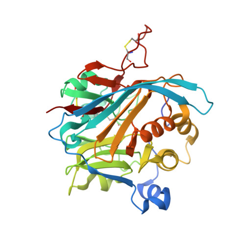
Crystal Structure of the Mannose-6-Phosphate Uncovering Enzyme.

Gorelik, A.Illes, K.Nagar, B.

(2020) Structure 28: 426-436.e3

  • DOI: https://doi.org/10.1016/j.str.2020.02.001
  • Primary Citation of Related Structures:  
    6PKG, 6PKH, 6PKI, 6PKU, 6PKY, 6U11

  • PubMed Abstract: 

    Most lysosomal hydrolytic enzymes reach their destination via the mannose-6-phosphate (M6P) pathway. The enzyme N-acetylglucosamine-1-phosphodiester α-N-acetylglucosaminidase (NAGPA, or "uncovering enzyme") catalyzes the second step in the M6P tag formation, namely the removal of the masking N-acetylglucosamine (GlcNAc) portion. Defects in this protein are associated with non-syndromic stuttering. To gain a better understanding of the function and regulation of this enzyme, we determined its crystal structure. The propeptide binds in a groove on the globular catalytic domain, blocking active site access. High-affinity substrate binding is enabled by a conformational switch in an active site loop. The protein recognizes the GlcNAc and phosphate portions of its substrate, but not the mannose moiety of the glycan. Based on enzymatic and 1 H-NMR analysis, a catalytic mechanism is proposed. Crystallographic and solution scattering analyses suggest that the C-terminal domain forms a long flexible stem that extends the enzyme away from the Golgi membrane.


  • Organizational Affiliation

    Department of Biochemistry, McGill University, Room 464, 3649 Promenade Sir-William-Osler, Montreal, QC H3G 0B1, Canada.


Macromolecules
Find similar proteins by:  (by identity cutoff)  |  3D Structure
Entity ID: 1
MoleculeChains Sequence LengthOrganismDetailsImage
N-acetylglucosamine-1-phosphodiester alpha-N-acetylglucosaminidase
A, B, C, D
309Danio rerioMutation(s): 2 
Gene Names: nagpa
UniProt
Find proteins for F1QSF9 (Danio rerio)
Explore F1QSF9 
Go to UniProtKB:  F1QSF9
Entity Groups  
Sequence Clusters30% Identity50% Identity70% Identity90% Identity95% Identity100% Identity
UniProt GroupF1QSF9
Sequence Annotations
Expand
  • Reference Sequence
Small Molecules
Ligands 5 Unique
IDChains Name / Formula / InChI Key2D Diagram3D Interactions
M6P (Subject of Investigation/LOI)
Query on M6P

Download Ideal Coordinates CCD File 
H [auth A],
N [auth B],
T [auth C],
U [auth C],
Z [auth D]
6-O-phosphono-alpha-D-mannopyranose
C6 H13 O9 P
NBSCHQHZLSJFNQ-PQMKYFCFSA-N
1PE
Query on 1PE

Download Ideal Coordinates CCD File 
I [auth A]PENTAETHYLENE GLYCOL
C10 H22 O6
JLFNLZLINWHATN-UHFFFAOYSA-N
NDG (Subject of Investigation/LOI)
Query on NDG

Download Ideal Coordinates CCD File 
G [auth A],
M [auth B],
S [auth C],
Y [auth D]
2-acetamido-2-deoxy-alpha-D-glucopyranose
C8 H15 N O6
OVRNDRQMDRJTHS-PVFLNQBWSA-N
NAG
Query on NAG

Download Ideal Coordinates CCD File 
E [auth A]
F [auth A]
K [auth B]
L [auth B]
Q [auth C]
E [auth A],
F [auth A],
K [auth B],
L [auth B],
Q [auth C],
R [auth C],
W [auth D],
X [auth D]
2-acetamido-2-deoxy-beta-D-glucopyranose
C8 H15 N O6
OVRNDRQMDRJTHS-FMDGEEDCSA-N
CL
Query on CL

Download Ideal Coordinates CCD File 
J [auth A],
O [auth B],
P [auth B],
V [auth C]
CHLORIDE ION
Cl
VEXZGXHMUGYJMC-UHFFFAOYSA-M
Experimental Data & Validation

Experimental Data

  • Method: X-RAY DIFFRACTION
  • Resolution: 2.29 Å
  • R-Value Free: 0.204 
  • R-Value Work: 0.170 
  • R-Value Observed: 0.171 
  • Space Group: P 41 21 2
Unit Cell:
Length ( Å )Angle ( ˚ )
a = 124.17α = 90
b = 124.17β = 90
c = 276.989γ = 90
Software Package:
Software NamePurpose
PHENIXrefinement
HKL-2000data reduction
HKL-2000data scaling
PHASERphasing

Structure Validation

View Full Validation Report


Ligand Structure Quality Assessment 


Entry History & Funding Information

Deposition Data


Funding OrganizationLocationGrant Number
Canadian Institutes of Health Research (CIHR)CanadaMOP-133535

Revision History  (Full details and data files)

  • Version 1.0: 2020-02-19
    Type: Initial release
  • Version 1.1: 2020-03-11
    Changes: Database references
  • Version 1.2: 2020-04-15
    Changes: Database references
  • Version 2.0: 2020-07-29
    Type: Remediation
    Reason: Carbohydrate remediation
    Changes: Atomic model, Data collection, Derived calculations, Structure summary
  • Version 2.1: 2023-10-11
    Changes: Data collection, Database references, Refinement description, Structure summary