6P3W

Crystal structure of the Cyclin A-CDK2-ORC1 complex


Experimental Data Snapshot

  • Method: X-RAY DIFFRACTION
  • Resolution: 2.54 Å
  • R-Value Free: 0.237 
  • R-Value Work: 0.198 
  • R-Value Observed: 0.200 

wwPDB Validation   3D Report Full Report


This is version 1.3 of the entry. See complete history


Literature

Structural basis for the ORC1-Cyclin A association.

Wang, B.Song, J.

(2019) Protein Sci 28: 1727-1733

  • DOI: https://doi.org/10.1002/pro.3689
  • Primary Citation of Related Structures:  
    6P3W

  • PubMed Abstract: 

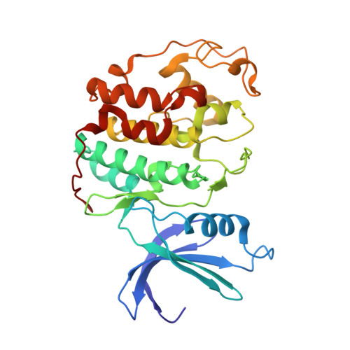
    Progression of cell cycle is regulated by sequential expression of cyclins, which associate with distinct cyclin kinases to drive the transition between different cell cycle phases. The complex of Cyclin A with cyclin-dependent kinase 2 (CDK2) controls the DNA replication activity through phosphorylation of a set of chromatin factors, which critically influences the S phase transition. It has been shown that the direct interaction between the Cyclin A-CDK2 complex and origin recognition complex subunit 1 (ORC1) mediates the localization of ORC1 to centrosomes, where ORC1 inhibits cyclin E-mediated centrosome reduplication. However, the molecular basis underlying the specific recognition between ORC1 and cyclins remains elusive. Here we report the crystal structure of Cyclin A-CDK2 complex bound to a peptide derived from ORC1 at 2.54 å resolution. The structure revealed that the ORC1 peptide interacts with a hydrophobic groove, termed cyclin binding groove (CBG), of Cyclin A via a KXL motif. Distinct from other identified CBG-binding sequences, an arginine residue flanking the KXL motif of ORC1 inserts into a neighboring acidic pocket, contributing to the strong ORC1-Cyclin A association. Furthermore, structural and sequence analysis of cyclins reveals divergence on the ORC1-binding sites, which may underpin their differential ORC1-binding activities. This study provides a structural basis of the specific ORC1-cyclins recognition, with implication in development of novel inhibitors against the cyclin/CDK complexes.


  • Organizational Affiliation

    Department of Biochemistry, University of California, Riverside, California.


Macromolecules
Find similar proteins by:  (by identity cutoff)  |  3D Structure
Entity ID: 1
MoleculeChains Sequence LengthOrganismDetailsImage
Cyclin-dependent kinase 2A,
D [auth C]
298Homo sapiensMutation(s): 0 
Gene Names: CDK2CDKN2
EC: 2.7.11.22
UniProt & NIH Common Fund Data Resources
Find proteins for P24941 (Homo sapiens)
Explore P24941 
Go to UniProtKB:  P24941
PHAROS:  P24941
GTEx:  ENSG00000123374 
Entity Groups  
Sequence Clusters30% Identity50% Identity70% Identity90% Identity95% Identity100% Identity
UniProt GroupP24941
Sequence Annotations
Expand
  • Reference Sequence
Find similar proteins by:  (by identity cutoff)  |  3D Structure
Entity ID: 2
MoleculeChains Sequence LengthOrganismDetailsImage
Cyclin-A2B,
E [auth D]
257Homo sapiensMutation(s): 0 
Gene Names: CCNA2CCN1CCNA
UniProt & NIH Common Fund Data Resources
Find proteins for P20248 (Homo sapiens)
Explore P20248 
Go to UniProtKB:  P20248
PHAROS:  P20248
GTEx:  ENSG00000145386 
Entity Groups  
Sequence Clusters30% Identity50% Identity70% Identity90% Identity95% Identity100% Identity
UniProt GroupP20248
Sequence Annotations
Expand
  • Reference Sequence

Find similar proteins by:  Sequence   |   3D Structure  

Entity ID: 3
MoleculeChains Sequence LengthOrganismDetailsImage
ORC1 PeptideC [auth E],
F
7Homo sapiensMutation(s): 0 
UniProt & NIH Common Fund Data Resources
Find proteins for Q13415 (Homo sapiens)
Explore Q13415 
Go to UniProtKB:  Q13415
PHAROS:  Q13415
GTEx:  ENSG00000085840 
Entity Groups  
UniProt GroupQ13415
Sequence Annotations
Expand
  • Reference Sequence
Small Molecules
Ligands 1 Unique
IDChains Name / Formula / InChI Key2D Diagram3D Interactions
PO4
Query on PO4

Download Ideal Coordinates CCD File 
G [auth A]
H [auth A]
I [auth A]
J [auth B]
K [auth C]
G [auth A],
H [auth A],
I [auth A],
J [auth B],
K [auth C],
L [auth C],
M [auth C],
N [auth C],
O [auth D],
P [auth D],
Q [auth D],
R [auth D]
PHOSPHATE ION
O4 P
NBIIXXVUZAFLBC-UHFFFAOYSA-K
Experimental Data & Validation

Experimental Data

  • Method: X-RAY DIFFRACTION
  • Resolution: 2.54 Å
  • R-Value Free: 0.237 
  • R-Value Work: 0.198 
  • R-Value Observed: 0.200 
  • Space Group: P 62 2 2
Unit Cell:
Length ( Å )Angle ( ˚ )
a = 186.396α = 90
b = 186.396β = 90
c = 214.707γ = 120
Software Package:
Software NamePurpose
PHENIXrefinement
HKL-2000data reduction
HKL-2000data scaling
PHASERphasing

Structure Validation

View Full Validation Report


Entry History 

Deposition Data

Revision History  (Full details and data files)

  • Version 1.0: 2019-07-31
    Type: Initial release
  • Version 1.1: 2019-08-28
    Changes: Data collection, Database references
  • Version 1.2: 2024-03-13
    Changes: Advisory, Data collection, Database references
  • Version 1.3: 2024-04-03
    Changes: Refinement description