6P25

Structure of S. cerevisiae protein O-mannosyltransferase Pmt1-Pmt2 complex bound to the sugar donor and a peptide acceptor


Experimental Data Snapshot

  • Method: ELECTRON MICROSCOPY
  • Resolution: 3.20 Å
  • Aggregation State: PARTICLE 
  • Reconstruction Method: SINGLE PARTICLE 

wwPDB Validation   3D Report Full Report


This is version 1.4 of the entry. See complete history


Literature

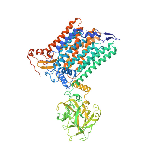
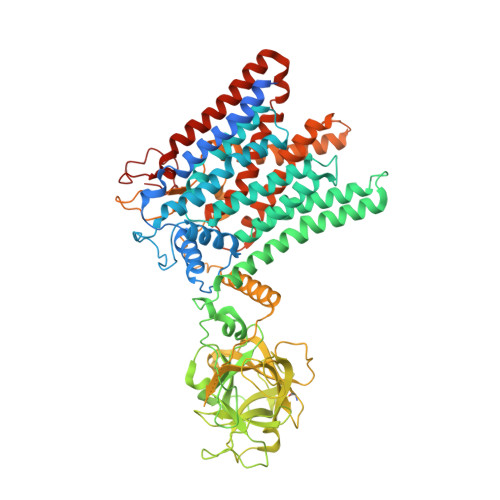
Structure of the eukaryotic protein O-mannosyltransferase Pmt1-Pmt2 complex.

Bai, L.Kovach, A.You, Q.Kenny, A.Li, H.

(2019) Nat Struct Mol Biol 26: 704-711

  • DOI: https://doi.org/10.1038/s41594-019-0262-6
  • Primary Citation of Related Structures:  
    6P25, 6P28, 6P2R

  • PubMed Abstract: 

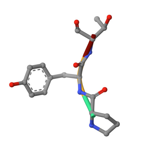
    In eukaryotes, a nascent peptide entering the endoplasmic reticulum (ER) is scanned by two Sec61 translocon-associated large membrane machines for protein N-glycosylation and protein O-mannosylation, respectively. While the structure of the eight-protein oligosaccharyltransferase complex has been determined recently, the structures of mannosyltransferases of the PMT family, which are an integral part of ER protein homeostasis, are still unknown. Here we report cryo-EM structures of the Saccharomyces cerevisiae Pmt1-Pmt2 complex bound to a donor and an acceptor peptide at 3.2-Å resolution, showing that each subunit contains 11 transmembrane helices and a lumenal β-trefoil fold termed the MIR domain. The structures reveal the substrate recognition model and confirm an inverting mannosyl-transferring reaction mechanism by the enzyme complex. Furthermore, we found that the transmembrane domains of Pmt1 and Pmt2 share a structural fold with the catalytic subunits of oligosaccharyltransferases, confirming a previously proposed evolutionary relationship between protein O-mannosylation and protein N-glycosylation.


  • Organizational Affiliation

    Structural Biology Program, Van Andel Research Institute, Grand Rapids, MI, USA.


Macromolecules
Find similar proteins by:  (by identity cutoff)  |  3D Structure
Entity ID: 1
MoleculeChains Sequence LengthOrganismDetailsImage
Dolichyl-phosphate-mannose--protein mannosyltransferase 1817Saccharomyces cerevisiae W303Mutation(s): 0 
Gene Names: PMT1YDL095WD2390
EC: 2.4.1.109
Membrane Entity: Yes 
UniProt
Find proteins for P33775 (Saccharomyces cerevisiae (strain ATCC 204508 / S288c))
Explore P33775 
Go to UniProtKB:  P33775
Entity Groups  
Sequence Clusters30% Identity50% Identity70% Identity90% Identity95% Identity100% Identity
UniProt GroupP33775
Sequence Annotations
Expand
  • Reference Sequence
Find similar proteins by:  (by identity cutoff)  |  3D Structure
Entity ID: 2
MoleculeChains Sequence LengthOrganismDetailsImage
Dolichyl-phosphate-mannose--protein mannosyltransferase 2759Saccharomyces cerevisiae W303Mutation(s): 0 
Gene Names: PMT2FUN25YAL023C
EC: 2.4.1.109
Membrane Entity: Yes 
UniProt
Find proteins for P31382 (Saccharomyces cerevisiae (strain ATCC 204508 / S288c))
Explore P31382 
Go to UniProtKB:  P31382
Entity Groups  
Sequence Clusters30% Identity50% Identity70% Identity90% Identity95% Identity100% Identity
UniProt GroupP31382
Sequence Annotations
Expand
  • Reference Sequence

Find similar proteins by:  Sequence   |   3D Structure  

Entity ID: 3
MoleculeChains Sequence LengthOrganismDetailsImage
acceptor peptideC [auth D]4Saccharomyces cerevisiae W303Mutation(s): 0 
Sequence Annotations
Expand
  • Reference Sequence
Experimental Data & Validation

Experimental Data

  • Method: ELECTRON MICROSCOPY
  • Resolution: 3.20 Å
  • Aggregation State: PARTICLE 
  • Reconstruction Method: SINGLE PARTICLE 
EM Software:
TaskSoftware PackageVersion
MODEL REFINEMENTPHENIX
MODEL REFINEMENTCoot
RECONSTRUCTIONCoot
RECONSTRUCTIONUCSF Chimera

Structure Validation

View Full Validation Report


Entry History & Funding Information

Deposition Data

  • Released Date: 2019-07-10 
  • Deposition Author(s): Bai, L., Li, H.

Funding OrganizationLocationGrant Number
National Institutes of Health/National Cancer Institute (NIH/NCI)United StatesR01-CA231466

Revision History  (Full details and data files)

  • Version 1.0: 2019-07-10
    Type: Initial release
  • Version 1.1: 2019-07-24
    Changes: Data collection, Database references
  • Version 1.2: 2019-08-21
    Changes: Data collection, Database references
  • Version 1.3: 2019-12-04
    Changes: Author supporting evidence
  • Version 1.4: 2020-07-29
    Type: Remediation
    Reason: Carbohydrate remediation
    Changes: Data collection, Derived calculations, Structure summary