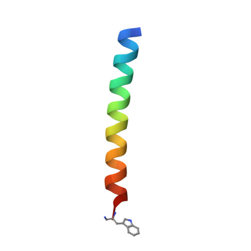
Retention of Coiled-Coil Dimer Formation in the Absence of Ion Pairing at Positions Flanking the Hydrophobic Core.
Biok, N.A., Passow, A.D., Wang, C., Bingman, C.A., Abbott, N.L., Gellman, S.H.(2019) Biochemistry 58: 4821-4826
- PubMed: 31738525
- DOI: https://doi.org/10.1021/acs.biochem.9b00668
- Primary Citation of Related Structures:
6OWD - PubMed Abstract:
Hydrophobic interactions govern how proteins fold and interact with other molecules, but the impact of nearby polar functionality on the effective hydrophobicity of nonpolar surfaces remains unclear. Here we use a common protein quaternary structure motif, the parallel coiled-coil dimer, to ask whether the identity of basic residues (arginine vs lysine; guanidinium vs ammonium) arrayed along one side of the constituent α-helices influences the favorability of dimerization driven by burial of hydrophobic surface on the other side of each helix. Significant sequence redesign was necessary to achieve the desired juxtaposition of nonpolar and cationic functionality, because we needed to eliminate charged side chains from positions flanking the nonpolar helix surface. Natural and designed sequences that form coiled coils are almost universally rich in acidic and basic residues at these flanking positions. Our arginine coiled-coil dimer was moderately more stable than the lysine analogue, which contrasts with behavior previously observed with helical β-amino acid oligomers bearing guanidinium versus ammonium groups. We attribute this backbone-dependent difference to variations in the extent to which the helical propensities of α- and β-residues can be modulated by design. These findings highlight the challenge of identifying noncovalent forces that direct structure formed by a flexible backbone.
Organizational Affiliation:
Department of Chemistry , University of Wisconsin-Madison , 1101 University Avenue , Madison , Wisconsin 53706 , United States.