6OOR

Structure of 1B1 bound to mouse CD1d


Experimental Data Snapshot

  • Method: X-RAY DIFFRACTION
  • Resolution: 2.45 Å
  • R-Value Free: 0.244 
  • R-Value Work: 0.208 
  • R-Value Observed: 0.209 

wwPDB Validation   3D Report Full Report

Currently 6OOR does not have a validation slider image.


This is version 1.4 of the entry. See complete history


Literature

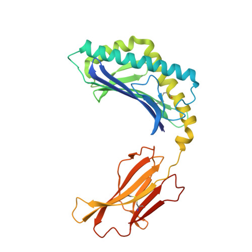
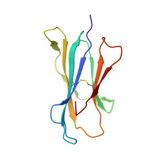
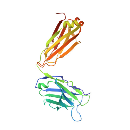
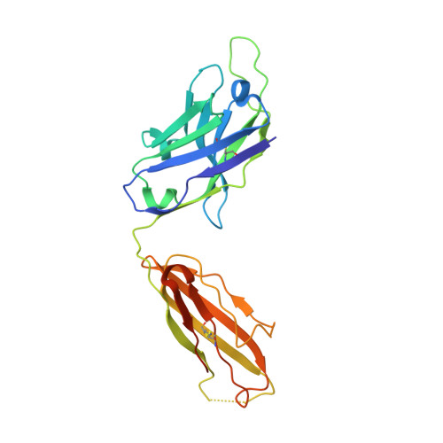
Structural basis of NKT cell inhibition using the T-cell receptor-blocking anti-CD1d antibody 1B1.

Ying, G.Wang, J.Mallevaey, T.Van Calenbergh, S.Zajonc, D.M.

(2019) J Biol Chem 294: 12947-12956

  • DOI: https://doi.org/10.1074/jbc.RA119.009403
  • Primary Citation of Related Structures:  
    6OOR

  • PubMed Abstract: 

    Natural killer T (NKT) cells are a subset of T lymphocytes that recognize glycolipid antigens presented by the CD1d molecule (CD1d). They rapidly respond to antigen challenge and can activate both innate and adaptive immune cells. To study the role of antigen presentation in NKT cell activation, previous studies have developed several anti-CD1d antibodies that block CD1d binding to T-cell receptors (TCRs). Antibodies that are specific to both CD1d and the presented antigen can only be used to study the function of only a limited number of antigens. In contrast, antibodies that bind CD1d and block TCR binding regardless of the presented antigen can be widely used to assess the role of TCR-mediated NKT cell activation in various disease models. Here, we report the crystal structure of the widely used anti-mouse CD1d antibody 1B1 bound to CD1d at a resolution of 2.45 Å and characterized its binding to CD1d-presented glycolipids. We observed that 1B1 uses a long hydrophobic H3 loop that is inserted deep into the binding groove of CD1d where it makes intimate nonpolar contacts with the lipid backbone of an incorporated spacer lipid. Using an NKT cell agonist that has a modified sphingosine moiety, we further demonstrate that 1B1 in its monovalent form cannot block TCR-mediated NKT cell activation, because 1B1 fails to bind with high affinity to mCD1d. Our results suggest potential limitations of using 1B1 to assess antigen recognition by NKT cells, especially when investigating antigens that do not follow the canonical two alkyl-chain rule.


  • Organizational Affiliation

    Division of Immune Regulation, La Jolla Institute for Immunology, La Jolla, California 92037.


Macromolecules
Find similar proteins by:  (by identity cutoff)  |  3D Structure
Entity ID: 1
MoleculeChains Sequence LengthOrganismDetailsImage
Antigen-presenting glycoprotein CD1d1285Mus musculusMutation(s): 1 
Gene Names: Cd1d1mCG_3074
UniProt & NIH Common Fund Data Resources
Find proteins for P11609 (Mus musculus)
Explore P11609 
Go to UniProtKB:  P11609
IMPC:  MGI:107674
Entity Groups  
Sequence Clusters30% Identity50% Identity70% Identity90% Identity95% Identity100% Identity
UniProt GroupP11609
Sequence Annotations
Expand
  • Reference Sequence
Find similar proteins by:  (by identity cutoff)  |  3D Structure
Entity ID: 2
MoleculeChains Sequence LengthOrganismDetailsImage
Beta-2-microglobulin99Mus musculusMutation(s): 0 
Gene Names: B2m
UniProt & NIH Common Fund Data Resources
Find proteins for P01887 (Mus musculus)
Explore P01887 
Go to UniProtKB:  P01887
IMPC:  MGI:88127
Entity Groups  
Sequence Clusters30% Identity50% Identity70% Identity90% Identity95% Identity100% Identity
UniProt GroupP01887
Sequence Annotations
Expand
  • Reference Sequence
Find similar proteins by:  (by identity cutoff)  |  3D Structure
Entity ID: 3
MoleculeChains Sequence LengthOrganismDetailsImage
Antibody 1B1 Light chainC [auth L]219Rattus norvegicusMutation(s): 0 
UniProt
Find proteins for A0A0G2JZW1 (Rattus norvegicus)
Explore A0A0G2JZW1 
Go to UniProtKB:  A0A0G2JZW1
Entity Groups  
Sequence Clusters30% Identity50% Identity70% Identity90% Identity95% Identity100% Identity
UniProt GroupA0A0G2JZW1
Sequence Annotations
Expand
  • Reference Sequence
Find similar proteins by:  (by identity cutoff)  |  3D Structure
Entity ID: 4
MoleculeChains Sequence LengthOrganismDetailsImage
Antibody 1B1 Heavy chainD [auth H]234Rattus norvegicusMutation(s): 0 
Entity Groups  
Sequence Clusters30% Identity50% Identity70% Identity90% Identity95% Identity100% Identity
Sequence Annotations
Expand
  • Reference Sequence
Experimental Data & Validation

Experimental Data

  • Method: X-RAY DIFFRACTION
  • Resolution: 2.45 Å
  • R-Value Free: 0.244 
  • R-Value Work: 0.208 
  • R-Value Observed: 0.209 
  • Space Group: C 2 2 21
Unit Cell:
Length ( Å )Angle ( ˚ )
a = 84.051α = 90
b = 160.964β = 90
c = 165.422γ = 90
Software Package:
Software NamePurpose
REFMACrefinement
HKL-2000data reduction
SCALEPACKdata scaling
PHASERphasing
PDB_EXTRACTdata extraction

Structure Validation

View Full Validation Report

Currently 6OOR does not have a validation slider image.


Entry History & Funding Information

Deposition Data


Funding OrganizationLocationGrant Number
National Institutes of Health/National Institute Of Allergy and Infectious Diseases (NIH/NIAID)United StatesAI137230

Revision History  (Full details and data files)

  • Version 1.0: 2019-07-17
    Type: Initial release
  • Version 1.1: 2019-07-24
    Changes: Data collection, Database references
  • Version 1.2: 2019-09-11
    Changes: Data collection, Database references
  • Version 1.3: 2019-12-18
    Changes: Author supporting evidence
  • Version 1.4: 2023-10-11
    Changes: Data collection, Database references, Derived calculations, Refinement description