


Crystal Structure of Dihydropteroate synthase from Mycobacterium smegmatis with bound 6-hydroxymethylpterin-monophosphate
Bolejack, M.J., Dranow, D.M., Lorimer, D.D., Horanyi, P.S., Edwards, T.E.To be published.
Experimental Data Snapshot
Entity ID: 1 | |||||
|---|---|---|---|---|---|
| Molecule | Chains | Sequence Length | Organism | Details | Image |
| Dihydropteroate synthase | 293 | Mycolicibacterium smegmatis MKD8 | Mutation(s): 1 Gene Names: D806_059810 EC: 2.5.1.15 | 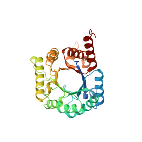 | |
UniProt | |||||
Find proteins for A0A2U9PYL8 (Mycolicibacterium smegmatis (strain MKD8)) Explore A0A2U9PYL8 Go to UniProtKB: A0A2U9PYL8 | |||||
Entity Groups | |||||
| Sequence Clusters | 30% Identity50% Identity70% Identity90% Identity95% Identity100% Identity | ||||
| UniProt Group | A0A2U9PYL8 | ||||
Sequence AnnotationsExpand | |||||
| |||||
| Ligands 4 Unique | |||||
|---|---|---|---|---|---|
| ID | Chains | Name / Formula / InChI Key | 2D Diagram | 3D Interactions | |
| PMM Query on PMM | D [auth A] | PTERIN-6-YL-METHYL-MONOPHOSPHATE C7 H8 N5 O5 P AJXFJEHKGGCFNM-UHFFFAOYSA-N |  | ||
| CIT Query on CIT | B [auth A], E [auth A] | CITRIC ACID C6 H8 O7 KRKNYBCHXYNGOX-UHFFFAOYSA-N |  | ||
| DMS Query on DMS | C [auth A] | DIMETHYL SULFOXIDE C2 H6 O S IAZDPXIOMUYVGZ-UHFFFAOYSA-N |  | ||
| EDO Query on EDO | F [auth A] G [auth A] H [auth A] I [auth A] J [auth A] | 1,2-ETHANEDIOL C2 H6 O2 LYCAIKOWRPUZTN-UHFFFAOYSA-N |  | ||
| Length ( Å ) | Angle ( ˚ ) |
|---|---|
| a = 47.72 | α = 90 |
| b = 96.41 | β = 90 |
| c = 135.07 | γ = 90 |
| Software Name | Purpose |
|---|---|
| PHENIX | refinement |
| XDS | data reduction |
| XSCALE | data scaling |
| PDB_EXTRACT | data extraction |
| MoRDa | phasing |
RCSB PDB (citation) is hosted by
RCSB PDB is a member of the

