6OEC

Yeast Spc42 Trimeric Coiled-Coil Amino Acids 181-211 fused to PDB: 3H5I


Experimental Data Snapshot

  • Method: X-RAY DIFFRACTION
  • Resolution: 2.51 Å
  • R-Value Free: 0.249 
  • R-Value Work: 0.195 
  • R-Value Observed: 0.198 

wwPDB Validation   3D Report Full Report


This is version 1.3 of the entry. See complete history


Literature

Structure and function of Spc42 coiled-coils in yeast centrosome assembly and duplication.

Drennan, A.C.Krishna, S.Seeger, M.A.Andreas, M.P.Gardner, J.M.Sether, E.K.R.Jaspersen, S.L.Rayment, I.

(2019) Mol Biol Cell 30: 1505-1522

  • DOI: https://doi.org/10.1091/mbc.E19-03-0167
  • Primary Citation of Related Structures:  
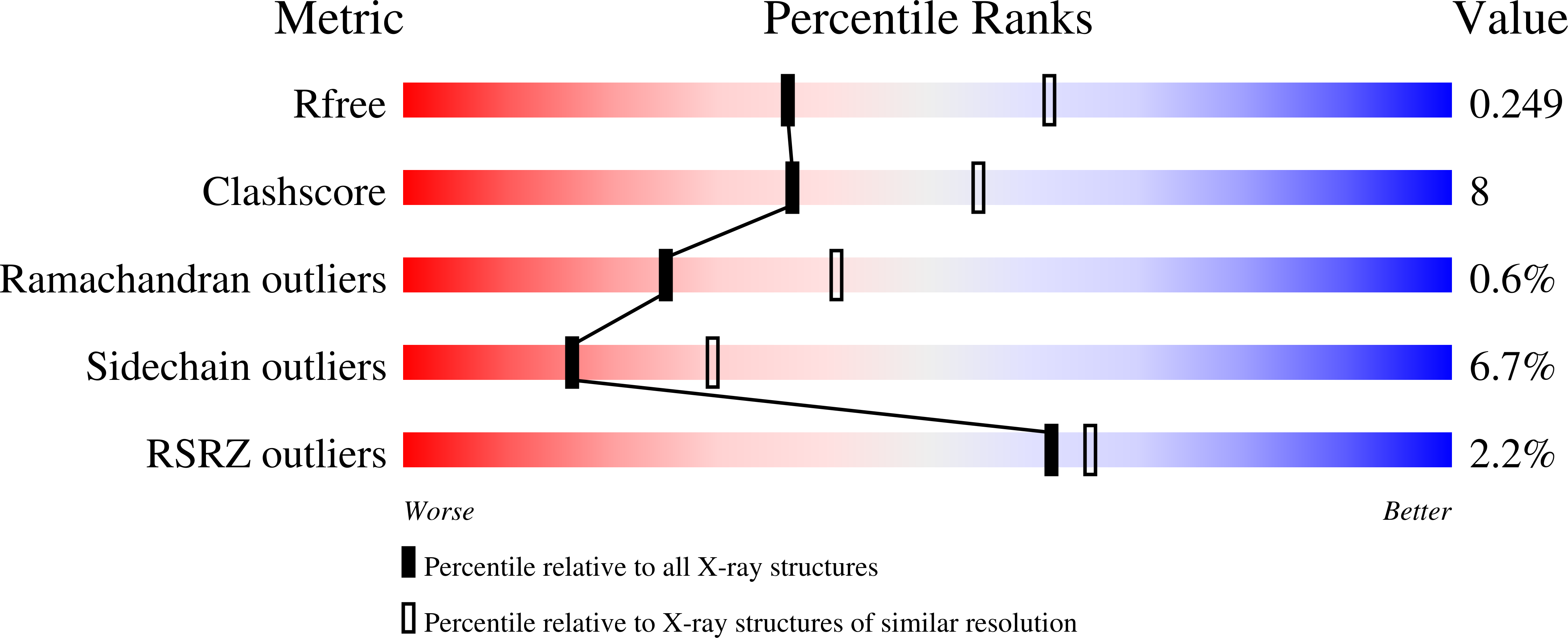
    6OD2, 6OEC, 6OEI

  • PubMed Abstract: 

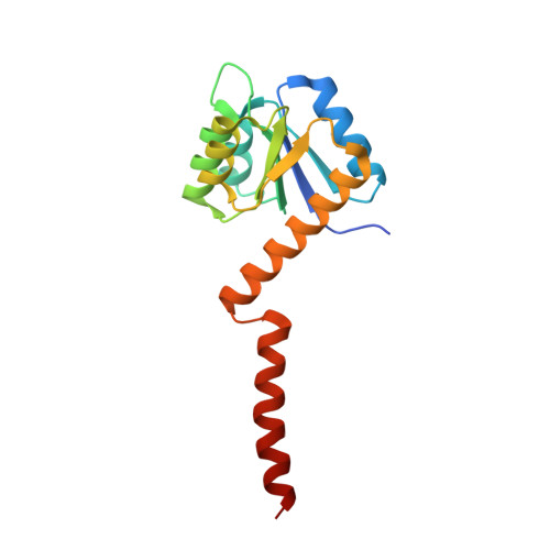
    Centrosomes and spindle pole bodies (SPBs) are membraneless organelles whose duplication and assembly is necessary for bipolar mitotic spindle formation. The structural organization and functional roles of major proteins in these organelles can provide critical insights into cell division control. Spc42, a phosphoregulated protein with an N-terminal dimeric coiled-coil (DCC), assembles into a hexameric array at the budding yeast SPB core, where it functions as a scaffold for SPB assembly. Here, we present in vitro and in vivo data to elucidate the structural arrangement and biological roles of Spc42 elements. Crystal structures reveal details of two additional coiled-coils in Spc42: a central trimeric coiled-coil and a C-terminal antiparallel DCC. Contributions of the three Spc42 coiled-coils and adjacent undetermined regions to the formation of an ∼145 Å hexameric lattice in an in vitro lipid monolayer assay and to SPB duplication and assembly in vivo reveal structural and functional redundancy in Spc42 assembly. We propose an updated model that incorporates the inherent symmetry of these Spc42 elements into a lattice, and thereby establishes the observed sixfold symmetry. The implications of this model for the organization of the central SPB core layer are discussed.


  • Organizational Affiliation

    Department of Biochemistry, University of Wisconsin-Madison, WI 53706.


Macromolecules
Find similar proteins by:  (by identity cutoff)  |  3D Structure
Entity ID: 1
MoleculeChains Sequence LengthOrganismDetailsImage
Response regulator/sensory box protein/GGDEF domain protein,Spindle pole body component SPC42
A, B, C, D, E
A, B, C, D, E, F, G, H, I, J, K, L
162Carboxydothermus hydrogenoformans Z-2901Saccharomyces cerevisiae S288C
This entity is chimeric
Mutation(s): 2 
Gene Names: CHY_0880SPC42YKL042WYKL255
UniProt
Find proteins for P36094 (Saccharomyces cerevisiae (strain ATCC 204508 / S288c))
Explore P36094 
Go to UniProtKB:  P36094
Find proteins for Q3ADQ4 (Carboxydothermus hydrogenoformans (strain ATCC BAA-161 / DSM 6008 / Z-2901))
Explore Q3ADQ4 
Go to UniProtKB:  Q3ADQ4
Entity Groups  
Sequence Clusters30% Identity50% Identity70% Identity90% Identity95% Identity100% Identity
UniProt GroupsQ3ADQ4P36094
Sequence Annotations
Expand
  • Reference Sequence
Experimental Data & Validation

Experimental Data

  • Method: X-RAY DIFFRACTION
  • Resolution: 2.51 Å
  • R-Value Free: 0.249 
  • R-Value Work: 0.195 
  • R-Value Observed: 0.198 
  • Space Group: P 21 21 2
Unit Cell:
Length ( Å )Angle ( ˚ )
a = 133.197α = 90
b = 169.723β = 90
c = 122.028γ = 90
Software Package:
Software NamePurpose
PHENIXrefinement
HKL-2000data reduction
HKL-2000data scaling
PHASERphasing
PDB_EXTRACTdata extraction

Structure Validation

View Full Validation Report


Entry History & Funding Information

Deposition Data


Funding OrganizationLocationGrant Number
National Institutes of Health/National Institute of General Medical Sciences (NIH/NIGMS)United StatesP01GM105537

Revision History  (Full details and data files)

  • Version 1.0: 2019-04-24
    Type: Initial release
  • Version 1.1: 2019-06-12
    Changes: Data collection, Database references
  • Version 1.2: 2020-01-01
    Changes: Author supporting evidence
  • Version 1.3: 2023-10-11
    Changes: Data collection, Database references, Refinement description