Sudan Ebolavirus VP35-NP Crystal Structure Reveals a Potential Target for Pan-Filovirus Treatment.
Landeras-Bueno, S., Oda, S.I., Norris, M.J., Li Salie, Z., Guenaga, J., Wyatt, R.T., Saphire, E.O.(2019) mBio 10
- PubMed: 31337716
- DOI: https://doi.org/10.1128/mBio.00734-19
- Primary Citation of Related Structures:
6OAF - PubMed Abstract:
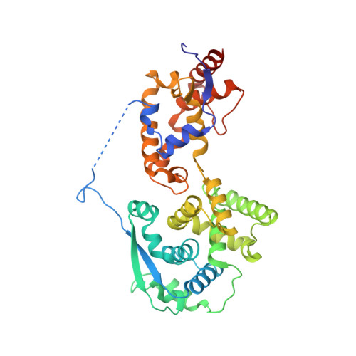
The filoviruses are etiological agents of life-threatening hemorrhagic fever with high mortality rate and risk of potential outbreak. Among members of this family, the Ebola (EBOV), Sudan (SUDV), and Marburg (MARV) viruses are considered the most pathogenic for humans. The ebolavirus nucleoprotein (NP) is the most abundant protein in infected cells and is essential for viral transcription and replication; thus, it represents an attractive target for therapeutic intervention. Here, we present the structure of SUDV NP in complex with the amino-terminal portion of the phosphoprotein VP35 at 2.3 Å. This structure captures VP35 chaperoning SUDV NP in a monomeric and RNA-free state. This transient state has been proposed to be key to maintaining a pool of monomeric and RNA-free NPs prior to NP-NP polymerization and encapsidation of the viral RNA genome. This structure also reveals a newly visualized interaction between NP and VP35, a well-defined beta sheet that is not present in previous structures. Affinity binding assays demonstrate that this beta sheet is essential for maintaining the high-affinity interaction between VP35 and a hydrophobic pocket on SUDV NP, and electron microscopy indicates the importance of this binding interaction to the oligomeric state and assembly of NP in human cells. Complementary structure-directed mutagenesis identifies critical residues conserved across the filovirus family that could be targeted by broadly effective antivirals. IMPORTANCE Outbreaks of the filoviruses can be unpredictable in timing, location, and identity of the causative virus, with each of Ebola virus, Sudan virus, Bundibugyo virus, and Marburg virus reemerging in the last several years to cause human disease with 30 to 90% lethality. The 2014-2016 outbreak in particular, with nearly 30,000 patients, highlighted the ability of these viruses to emerge unexpectedly and spread rapidly. Two ebolavirus outbreaks have emerged this year, yet we still lack FDA-approved drugs with pan-filovirus activity to treat existing and emergent ebolaviruses. For all filoviruses, the interaction between the nucleoprotein and the phosphoprotein is essential for the virus life cycle and is a potential target for therapeutic intervention. In this report, we describe the crystal structure of the SUDV nucleoprotein with the interacting domain of the viral phosphoprotein, and we identify residues critical for high-affinity interaction and for control of the oligomeric state of the nucleoprotein. Structural comparison of this heterodimer with other members of the filovirus family allowed us to find conserved and essential atomic features that will facilitate understanding of the virus life cycle and the rational design of antivirals.
Organizational Affiliation:
La Jolla Institute for Immunology, La Jolla, California, USA.