6OA9

Cdc48-Npl4 complex processing poly-ubiquitinated substrate in the presence of ATP


Experimental Data Snapshot

  • Method: ELECTRON MICROSCOPY
  • Resolution: 3.90 Å
  • Aggregation State: PARTICLE 
  • Reconstruction Method: SINGLE PARTICLE 

wwPDB Validation   3D Report Full Report


This is version 1.4 of the entry. See complete history


Literature

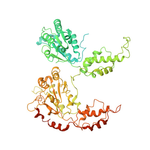
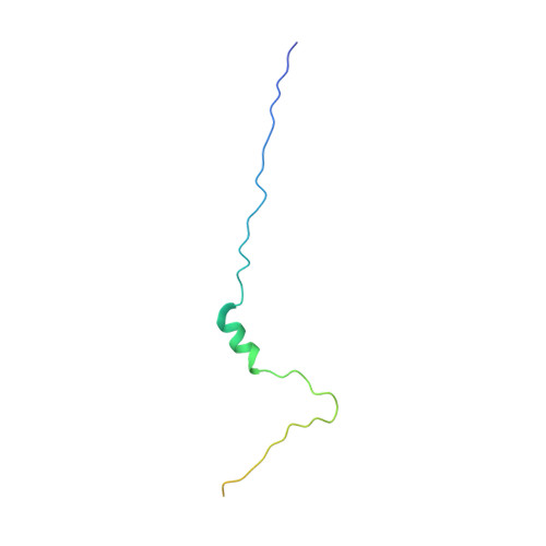
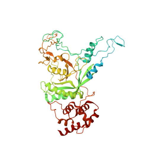
Substrate processing by the Cdc48 ATPase complex is initiated by ubiquitin unfolding.

Twomey, E.C.Ji, Z.Wales, T.E.Bodnar, N.O.Ficarro, S.B.Marto, J.A.Engen, J.R.Rapoport, T.A.

(2019) Science 365

  • DOI: https://doi.org/10.1126/science.aax1033
  • Primary Citation of Related Structures:  
    6OA9, 6OAA, 6OAB

  • PubMed Abstract: 

    The Cdc48 adenosine triphosphatase (ATPase) (p97 or valosin-containing protein in mammals) and its cofactor Ufd1/Npl4 extract polyubiquitinated proteins from membranes or macromolecular complexes for subsequent degradation by the proteasome. How Cdc48 processes its diverse and often well-folded substrates is unclear. Here, we report cryo-electron microscopy structures of the Cdc48 ATPase in complex with Ufd1/Npl4 and polyubiquitinated substrate. The structures show that the Cdc48 complex initiates substrate processing by unfolding a ubiquitin molecule. The unfolded ubiquitin molecule binds to Npl4 and projects its N-terminal segment through both hexameric ATPase rings. Pore loops of the second ring form a staircase that acts as a conveyer belt to move the polypeptide through the central pore. Inducing the unfolding of ubiquitin allows the Cdc48 ATPase complex to process a broad range of substrates.


  • Organizational Affiliation

    Department of Cell Biology, Harvard Medical School, and Howard Hughes Medical Institute, 240 Longwood Avenue, Boston, MA 02115, USA.


Macromolecules
Find similar proteins by:  (by identity cutoff)  |  3D Structure
Entity ID: 1
MoleculeChains Sequence LengthOrganismDetailsImage
Cell division control protein 48
A, B, C, D, E
A, B, C, D, E, F
835Saccharomyces cerevisiaeMutation(s): 0 
Gene Names: CDC48YDL126C
EC: 3.6.4.6
UniProt
Find proteins for P25694 (Saccharomyces cerevisiae (strain ATCC 204508 / S288c))
Explore P25694 
Go to UniProtKB:  P25694
Entity Groups  
Sequence Clusters30% Identity50% Identity70% Identity90% Identity95% Identity100% Identity
UniProt GroupP25694
Sequence Annotations
Expand
  • Reference Sequence
Find similar proteins by:  (by identity cutoff)  |  3D Structure
Entity ID: 2
MoleculeChains Sequence LengthOrganismDetailsImage
UbiquitinG [auth K],
H,
I [auth J]
76Saccharomyces cerevisiaeMutation(s): 0 
Gene Names: RPL40AUBI1YIL148W
UniProt
Find proteins for P0CH08 (Saccharomyces cerevisiae (strain ATCC 204508 / S288c))
Explore P0CH08 
Go to UniProtKB:  P0CH08
Entity Groups  
Sequence Clusters30% Identity50% Identity70% Identity90% Identity95% Identity100% Identity
UniProt GroupP0CH08
Sequence Annotations
Expand
  • Reference Sequence
Find similar proteins by:  (by identity cutoff)  |  3D Structure
Entity ID: 3
MoleculeChains Sequence LengthOrganismDetailsImage
Nuclear protein localization protein 4J [auth G]580Saccharomyces cerevisiaeMutation(s): 0 
Gene Names: NPL4HRD4YBR170CYBR1231
UniProt
Find proteins for P33755 (Saccharomyces cerevisiae (strain ATCC 204508 / S288c))
Explore P33755 
Go to UniProtKB:  P33755
Entity Groups  
Sequence Clusters30% Identity50% Identity70% Identity90% Identity95% Identity100% Identity
UniProt GroupP33755
Sequence Annotations
Expand
  • Reference Sequence
Small Molecules
Ligands 3 Unique
IDChains Name / Formula / InChI Key2D Diagram3D Interactions
ATP
Query on ATP

Download Ideal Coordinates CCD File 
K [auth A]
L [auth B]
M [auth C]
N [auth C]
O [auth C]
K [auth A],
L [auth B],
M [auth C],
N [auth C],
O [auth C],
P [auth D],
Q [auth D],
S [auth E],
T [auth F]
ADENOSINE-5'-TRIPHOSPHATE
C10 H16 N5 O13 P3
ZKHQWZAMYRWXGA-KQYNXXCUSA-N
ADP
Query on ADP

Download Ideal Coordinates CCD File 
R [auth E]ADENOSINE-5'-DIPHOSPHATE
C10 H15 N5 O10 P2
XTWYTFMLZFPYCI-KQYNXXCUSA-N
ZN
Query on ZN

Download Ideal Coordinates CCD File 
U [auth G],
V [auth G]
ZINC ION
Zn
PTFCDOFLOPIGGS-UHFFFAOYSA-N
Experimental Data & Validation

Experimental Data

  • Method: ELECTRON MICROSCOPY
  • Resolution: 3.90 Å
  • Aggregation State: PARTICLE 
  • Reconstruction Method: SINGLE PARTICLE 

Structure Validation

View Full Validation Report


Entry History & Funding Information

Deposition Data


Funding OrganizationLocationGrant Number
Howard Hughes Medical Institute (HHMI)United States--
National Institutes of Health/National Institute of General Medical Sciences (NIH/NIGMS)United StatesR01GM052586
National Institutes of Health/National Institute of General Medical Sciences (NIH/NIGMS)United StatesT32GM007753

Revision History  (Full details and data files)

  • Version 1.0: 2019-07-03
    Type: Initial release
  • Version 1.1: 2019-07-10
    Changes: Data collection, Database references
  • Version 1.2: 2019-08-14
    Changes: Data collection, Database references
  • Version 1.3: 2019-11-20
    Changes: Author supporting evidence
  • Version 1.4: 2024-03-20
    Changes: Data collection, Database references