6O8B

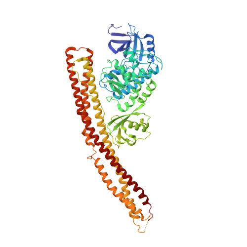
Crystal structure of STING CTD in complex with TBK1


Experimental Data Snapshot

  • Method: X-RAY DIFFRACTION
  • Resolution: 3.40 Å
  • R-Value Free: 0.258 
  • R-Value Work: 0.243 
  • R-Value Observed: 0.244 

wwPDB Validation   3D Report Full Report


Ligand Structure Quality Assessment 


This is version 1.3 of the entry. See complete history


Literature

A conserved PLPLRT/SD motif of STING mediates the recruitment and activation of TBK1.

Zhao, B.Du, F.Xu, P.Shu, C.Sankaran, B.Bell, S.L.Liu, M.Lei, Y.Gao, X.Fu, X.Zhu, F.Liu, Y.Laganowsky, A.Zheng, X.Ji, J.Y.West, A.P.Watson, R.O.Li, P.

(2019) Nature 569: 718-722

  • DOI: https://doi.org/10.1038/s41586-019-1228-x
  • Primary Citation of Related Structures:  
    6O8B, 6O8C

  • PubMed Abstract: 

    Nucleic acids from bacteria or viruses induce potent immune responses in infected cells 1-4 . The detection of pathogen-derived nucleic acids is a central strategy by which the host senses infection and initiates protective immune responses 5,6 . Cyclic GMP-AMP synthase (cGAS) is a double-stranded DNA sensor 7,8 . It catalyses the synthesis of cyclic GMP-AMP (cGAMP) 9-12 , which stimulates the induction of type I interferons through the STING-TBK1-IRF-3 signalling axis 13-15 . STING oligomerizes after binding of cGAMP, leading to the recruitment and activation of the TBK1 kinase 8,16 . The IRF-3 transcription factor is then recruited to the signalling complex and activated by TBK1 8,17-20 . Phosphorylated IRF-3 translocates to the nucleus and initiates the expression of type I interferons 21 . However, the precise mechanisms that govern activation of STING by cGAMP and subsequent activation of TBK1 by STING remain unclear. Here we show that a conserved PLPLRT/SD motif within the C-terminal tail of STING mediates the recruitment and activation of TBK1. Crystal structures of TBK1 bound to STING reveal that the PLPLRT/SD motif binds to the dimer interface of TBK1. Cell-based studies confirm that the direct interaction between TBK1 and STING is essential for induction of IFNβ after cGAMP stimulation. Moreover, we show that full-length STING oligomerizes after it binds cGAMP, and highlight this as an essential step in the activation of STING-mediated signalling. These findings provide a structural basis for the development of STING agonists and antagonists for the treatment of cancer and autoimmune disorders.


  • Organizational Affiliation

    Department of Biochemistry and Biophysics, Texas A&M University, College Station, TX, USA.


Macromolecules
Find similar proteins by:  (by identity cutoff)  |  3D Structure
Entity ID: 1
MoleculeChains Sequence LengthOrganismDetailsImage
Stimulator of interferon genes proteinA [auth D],
B [auth E]
226Homo sapiensMutation(s): 4 
Gene Names: TMEM173ERISMITASTING
UniProt & NIH Common Fund Data Resources
Find proteins for Q86WV6 (Homo sapiens)
Explore Q86WV6 
Go to UniProtKB:  Q86WV6
PHAROS:  Q86WV6
GTEx:  ENSG00000184584 
Entity Groups  
Sequence Clusters30% Identity50% Identity70% Identity90% Identity95% Identity100% Identity
UniProt GroupQ86WV6
Sequence Annotations
Expand
  • Reference Sequence
Find similar proteins by:  (by identity cutoff)  |  3D Structure
Entity ID: 2
MoleculeChains Sequence LengthOrganismDetailsImage
Serine/threonine-protein kinase TBK1C [auth A],
D [auth B]
665Homo sapiensMutation(s): 4 
Gene Names: TBK1NAK
EC: 2.7.11.1
UniProt & NIH Common Fund Data Resources
Find proteins for Q9UHD2 (Homo sapiens)
Explore Q9UHD2 
Go to UniProtKB:  Q9UHD2
PHAROS:  Q9UHD2
GTEx:  ENSG00000183735 
Entity Groups  
Sequence Clusters30% Identity50% Identity70% Identity90% Identity95% Identity100% Identity
UniProt GroupQ9UHD2
Sequence Annotations
Expand
  • Reference Sequence
Small Molecules
Ligands 1 Unique
IDChains Name / Formula / InChI Key2D Diagram3D Interactions
BX7
Query on BX7

Download Ideal Coordinates CCD File 
E [auth A],
F [auth B]
N-(3-{[5-iodo-4-({3-[(thiophen-2-ylcarbonyl)amino]propyl}amino)pyrimidin-2-yl]amino}phenyl)pyrrolidine-1-carboxamide
C23 H26 I N7 O2 S
VAVXGGRQQJZYBL-UHFFFAOYSA-N
Experimental Data & Validation

Experimental Data

  • Method: X-RAY DIFFRACTION
  • Resolution: 3.40 Å
  • R-Value Free: 0.258 
  • R-Value Work: 0.243 
  • R-Value Observed: 0.244 
  • Space Group: P 65 2 2
Unit Cell:
Length ( Å )Angle ( ˚ )
a = 250.69α = 90
b = 250.69β = 90
c = 239.241γ = 120
Software Package:
Software NamePurpose
PHENIXrefinement
iMOSFLMdata reduction
pointlessdata scaling
PHENIXphasing

Structure Validation

View Full Validation Report


Ligand Structure Quality Assessment 


Entry History & Funding Information

Deposition Data


Funding OrganizationLocationGrant Number
Welch FoundationUnited StatesA-1931-20170325

Revision History  (Full details and data files)

  • Version 1.0: 2019-05-22
    Type: Initial release
  • Version 1.1: 2019-06-05
    Changes: Data collection, Database references
  • Version 1.2: 2019-06-12
    Changes: Data collection, Database references
  • Version 1.3: 2023-10-11
    Changes: Data collection, Database references, Refinement description