6O4W

Binary complex of native hAChE with Donepezil


Experimental Data Snapshot

  • Method: X-RAY DIFFRACTION
  • Resolution: 2.35 Å
  • R-Value Free: 0.212 
  • R-Value Work: 0.186 
  • R-Value Observed: 0.187 

wwPDB Validation   3D Report Full Report


Ligand Structure Quality Assessment 


This is version 1.4 of the entry. See complete history


Literature

A new crystal form of human acetylcholinesterase for exploratory room-temperature crystallography studies.

Gerlits, O.Ho, K.Y.Cheng, X.Blumenthal, D.Taylor, P.Kovalevsky, A.Radic, Z.

(2019) Chem Biol Interact 309: 108698-108698

  • DOI: https://doi.org/10.1016/j.cbi.2019.06.011
  • Primary Citation of Related Structures:  
    6O4W, 6O4X, 6O50, 6O52

  • PubMed Abstract: 

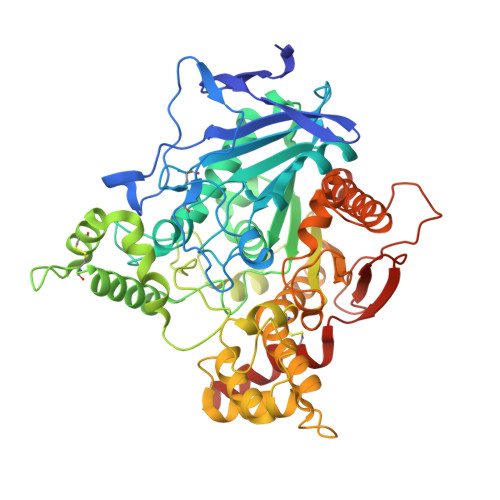
    Structure-guided design of novel pharmacologically active molecules relies at least in part on functionally relevant accuracy of macromolecular structures for template based drug design. Currently, about 95% of all macromolecular X-ray structures available in the PDB (Protein Data Bank) were obtained from diffraction experiments at low, cryogenic temperatures. However, it is known that functionally relevant conformations of both macromolecules and pharmacological ligands can differ at higher, physiological temperatures. We describe in this article development and properties of new human acetylcholinesterase (AChE) crystals of space group P3 1 and a new unit cell, amenable for room-temperature X-ray diffraction studies. We co-crystallized hAChE in P3 1 unit cell with the reversible inhibitor 9-aminoacridine that binds at the base of the active center gorge in addition to inhibitors that span the full length of the gorge, donepezil (Aricept, E2020) and AChE specific inhibitor BW284c51. Their new low temperature P3 1 space group structures appear similar to those previously obtained in the different P3 1 21 unit cell. Successful solution of the new room temperature 3.2 Å resolution structure of BW284c51*hAChE complex from large P3 1 crystals enables us to proceed with studying room temperature structures of lower affinity complexes, such as oxime reactivators bound to hAChE, where temperature-related conformational diversity could be expected in both oxime and hAChE, which could lead to better informed structure-based design under conditions approaching physiological temperature.


  • Organizational Affiliation

    (a)Bredesen Center, University of Tennessee, Knoxville, TN, 37996, USA.


Macromolecules
Find similar proteins by:  (by identity cutoff)  |  3D Structure
Entity ID: 1
MoleculeChains Sequence LengthOrganismDetailsImage
Acetylcholinesterase
A, B
550Homo sapiensMutation(s): 0 
Gene Names: ACHE
EC: 3.1.1.7
UniProt & NIH Common Fund Data Resources
Find proteins for P22303 (Homo sapiens)
Explore P22303 
Go to UniProtKB:  P22303
PHAROS:  P22303
GTEx:  ENSG00000087085 
Entity Groups  
Sequence Clusters30% Identity50% Identity70% Identity90% Identity95% Identity100% Identity
UniProt GroupP22303
Sequence Annotations
Expand
  • Reference Sequence
Small Molecules
Ligands 3 Unique
IDChains Name / Formula / InChI Key2D Diagram3D Interactions
E20
Query on E20

Download Ideal Coordinates CCD File 
F [auth A],
K [auth B]
1-BENZYL-4-[(5,6-DIMETHOXY-1-INDANON-2-YL)METHYL]PIPERIDINE
C24 H29 N O3
ADEBPBSSDYVVLD-HXUWFJFHSA-N
GOL
Query on GOL

Download Ideal Coordinates CCD File 
C [auth A]
D [auth A]
E [auth A]
H [auth B]
I [auth B]
C [auth A],
D [auth A],
E [auth A],
H [auth B],
I [auth B],
J [auth B]
GLYCEROL
C3 H8 O3
PEDCQBHIVMGVHV-UHFFFAOYSA-N
NO3
Query on NO3

Download Ideal Coordinates CCD File 
G [auth A],
L [auth B],
M [auth B],
N [auth B]
NITRATE ION
N O3
NHNBFGGVMKEFGY-UHFFFAOYSA-N
Binding Affinity Annotations 
IDSourceBinding Affinity
E20 BindingDB:  6O4W Ki: min: 2.9, max: 38 (nM) from 9 assay(s)
IC50: min: 0.12, max: 1060 (nM) from 73 assay(s)
EC50: 43 (nM) from 1 assay(s)
Experimental Data & Validation

Experimental Data

  • Method: X-RAY DIFFRACTION
  • Resolution: 2.35 Å
  • R-Value Free: 0.212 
  • R-Value Work: 0.186 
  • R-Value Observed: 0.187 
  • Space Group: P 31
Unit Cell:
Length ( Å )Angle ( ˚ )
a = 123.792α = 90
b = 123.792β = 90
c = 129.413γ = 120
Software Package:
Software NamePurpose
PHENIXrefinement
HKL-3000data reduction
HKL-3000data scaling
PHASERphasing

Structure Validation

View Full Validation Report


Ligand Structure Quality Assessment 


Entry History & Funding Information

Deposition Data


Funding OrganizationLocationGrant Number
National Institutes of Health/National Institute of Neurological Disorders and Stroke (NIH/NINDS)United States1U01NS083451

Revision History  (Full details and data files)

  • Version 1.0: 2019-06-19
    Type: Initial release
  • Version 1.1: 2019-07-10
    Changes: Data collection, Database references
  • Version 1.2: 2019-12-18
    Changes: Author supporting evidence
  • Version 1.3: 2020-02-19
    Changes: Derived calculations
  • Version 1.4: 2023-10-11
    Changes: Data collection, Database references, Refinement description