6NZK

Structural basis for human coronavirus attachment to sialic acid receptors


Experimental Data Snapshot

  • Method: ELECTRON MICROSCOPY
  • Resolution: 2.80 Å
  • Aggregation State: PARTICLE 
  • Reconstruction Method: SINGLE PARTICLE 

wwPDB Validation   3D Report Full Report


This is version 2.0 of the entry. See complete history


Literature

Structural basis for human coronavirus attachment to sialic acid receptors.

Alejandra Tortorici, M.Walls, A.C.Lang, Y.Wang, C.Li, Z.Koerhuis, D.Boons, G.J.Bosch, B.J.Rey, F.A.de Groot, R.J.Veesler, D.

(2019) Nat Struct Mol Biol 26: 481-489

  • DOI: https://doi.org/10.1038/s41594-019-0233-y
  • Primary Citation of Related Structures:  
    6NZK, 6OHW

  • PubMed Abstract: 

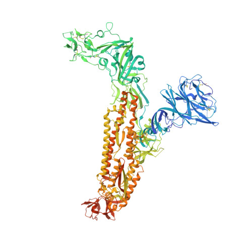
    Coronaviruses cause respiratory tract infections in humans and outbreaks of deadly pneumonia worldwide. Infections are initiated by the transmembrane spike (S) glycoprotein, which binds to host receptors and fuses the viral and cellular membranes. To understand the molecular basis of coronavirus attachment to oligosaccharide receptors, we determined cryo-EM structures of coronavirus OC43 S glycoprotein trimer in isolation and in complex with a 9-O-acetylated sialic acid. We show that the ligand binds with fast kinetics to a surface-exposed groove and that interactions at the identified site are essential for S-mediated viral entry into host cells, but free monosaccharide does not trigger fusogenic conformational changes. The receptor-interacting site is conserved in all coronavirus S glycoproteins that engage 9-O-acetyl-sialogycans, with an architecture similar to those of the ligand-binding pockets of coronavirus hemagglutinin esterases and influenza virus C/D hemagglutinin-esterase fusion glycoproteins. Our results demonstrate these viruses evolved similar strategies to engage sialoglycans at the surface of target cells.


  • Organizational Affiliation

    Department of Biochemistry, University of Washington, Seattle, WA, USA.


Macromolecules
Find similar proteins by:  (by identity cutoff)  |  3D Structure
Entity ID: 1
MoleculeChains Sequence LengthOrganismDetailsImage
Spike surface glycoprotein
A, B, C
1,322Human coronavirus OC43Mutation(s): 3 
UniProt
Find proteins for Q696P8 (Human coronavirus OC43)
Explore Q696P8 
Go to UniProtKB:  Q696P8
Entity Groups  
Sequence Clusters30% Identity50% Identity70% Identity90% Identity95% Identity100% Identity
UniProt GroupQ696P8
Sequence Annotations
Expand
  • Reference Sequence
Oligosaccharides

Help

Entity ID: 2
MoleculeChains Length2D Diagram Glycosylation3D Interactions
beta-D-mannopyranose-(1-4)-2-acetamido-2-deoxy-beta-D-glucopyranose-(1-4)-2-acetamido-2-deoxy-beta-D-glucopyranose
AA [auth a],
BA [auth b],
D,
E,
G,
AA [auth a],
BA [auth b],
D,
E,
G,
GA [auth g],
H,
M,
N,
O,
Q,
R,
W,
X,
Y
3N-Glycosylation
Glycosylation Resources
GlyTouCan:  G15407YE
GlyCosmos:  G15407YE
GlyGen:  G15407YE
Entity ID: 3
MoleculeChains Length2D Diagram Glycosylation3D Interactions
alpha-D-mannopyranose-(1-3)-beta-D-mannopyranose-(1-4)-2-acetamido-2-deoxy-beta-D-glucopyranose-(1-4)-2-acetamido-2-deoxy-beta-D-glucopyranose
DA [auth d],
F,
J,
P,
T,
DA [auth d],
F,
J,
P,
T,
Z
4N-Glycosylation
Glycosylation Resources
GlyTouCan:  G81315DD
GlyCosmos:  G81315DD
GlyGen:  G81315DD
Entity ID: 4
MoleculeChains Length2D Diagram Glycosylation3D Interactions
2-acetamido-2-deoxy-beta-D-glucopyranose-(1-4)-2-acetamido-2-deoxy-beta-D-glucopyranose
CA [auth c],
EA [auth e],
FA [auth f],
I,
K,
CA [auth c],
EA [auth e],
FA [auth f],
I,
K,
L,
S,
U,
V
2N-Glycosylation
Glycosylation Resources
GlyTouCan:  G42666HT
GlyCosmos:  G42666HT
GlyGen:  G42666HT
Small Molecules
Ligands 2 Unique
IDChains Name / Formula / InChI Key2D Diagram3D Interactions
MJJ
Query on MJJ

Download Ideal Coordinates CCD File 
MA [auth A],
SA [auth B],
YA [auth C]
methyl 9-O-acetyl-5-acetamido-3,5-dideoxy-D-glycero-alpha-D-galacto-non-2-ulopyranosidonic acid
C14 H23 N O10
NIEBVOWRRSQMQG-KXEMTNKZSA-N
NAG
Query on NAG

Download Ideal Coordinates CCD File 
HA [auth A]
IA [auth A]
JA [auth A]
KA [auth A]
LA [auth A]
HA [auth A],
IA [auth A],
JA [auth A],
KA [auth A],
LA [auth A],
NA [auth B],
OA [auth B],
PA [auth B],
QA [auth B],
RA [auth B],
TA [auth C],
UA [auth C],
VA [auth C],
WA [auth C],
XA [auth C]
2-acetamido-2-deoxy-beta-D-glucopyranose
C8 H15 N O6
OVRNDRQMDRJTHS-FMDGEEDCSA-N
Experimental Data & Validation

Experimental Data

  • Method: ELECTRON MICROSCOPY
  • Resolution: 2.80 Å
  • Aggregation State: PARTICLE 
  • Reconstruction Method: SINGLE PARTICLE 
EM Software:
TaskSoftware PackageVersion
RECONSTRUCTIONcryoSPARC

Structure Validation

View Full Validation Report


Entry History & Funding Information

Deposition Data


Funding OrganizationLocationGrant Number
National Institutes of Health/National Institute of General Medical Sciences (NIH/NIGMS)United StatesGM120553

Revision History  (Full details and data files)

  • Version 1.0: 2019-06-05
    Type: Initial release
  • Version 1.1: 2019-06-19
    Changes: Data collection, Data processing, Database references
  • Version 1.2: 2020-01-08
    Changes: Author supporting evidence
  • Version 2.0: 2020-07-29
    Type: Remediation
    Reason: Carbohydrate remediation
    Changes: Atomic model, Data collection, Derived calculations, Structure summary