6NK1

EphA2 LBD in complex with bA-WLA-YRPKbio peptide


Experimental Data Snapshot

  • Method: X-RAY DIFFRACTION
  • Resolution: 1.55 Å
  • R-Value Free: 0.169 
  • R-Value Work: 0.146 
  • R-Value Observed: 0.147 

wwPDB Validation   3D Report Full Report


This is version 1.4 of the entry. See complete history


Literature

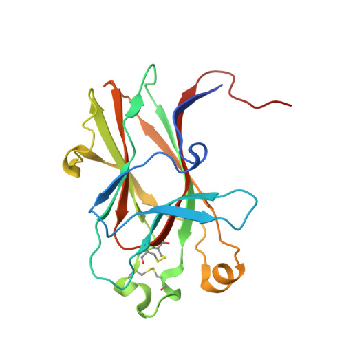
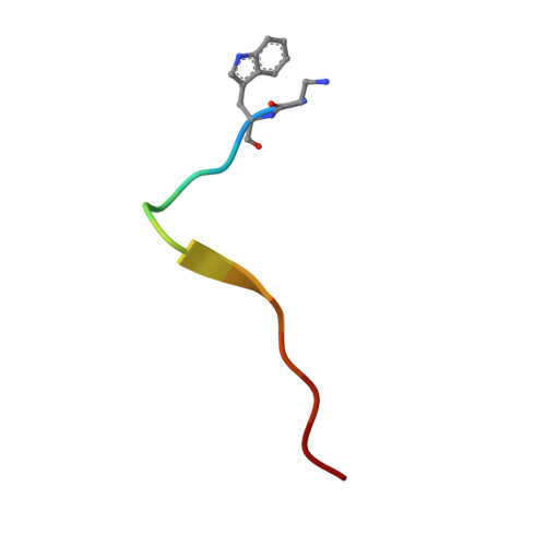
Engineering nanomolar peptide ligands that differentially modulate EphA2 receptor signaling.

Gomez-Soler, M.Petersen Gehring, M.Lechtenberg, B.C.Zapata-Mercado, E.Hristova, K.Pasquale, E.B.

(2019) J Biol Chem 294: 8791-8805

  • DOI: https://doi.org/10.1074/jbc.RA119.008213
  • Primary Citation of Related Structures:  
    6NJZ, 6NK0, 6NK1, 6NK2, 6NKP

  • PubMed Abstract: 

    The EPH receptor A2 (EphA2) tyrosine kinase plays an important role in a plethora of biological and disease processes, ranging from angiogenesis and cancer to inflammation and parasitic infections. EphA2 is therefore considered an important drug target. Two short peptides previously identified by phage display, named YSA and SWL, are widely used as EphA2-targeting agents owing to their high specificity for this receptor. However, these peptides have only modest (micromolar) potency. Lack of structural information on the binding interactions of YSA and SWL with the extracellular EphA2 ligand-binding domain (LBD) has for many years precluded structure-guided improvements. We now report the high-resolution (1.53-2.20 Å) crystal structures of the YSA peptide and several of its improved derivatives in complex with the EphA2 LBD, disclosing that YSA targets the ephrin-binding pocket of EphA2 and mimics binding features of the ephrin-A ligands. The structural information obtained enabled iterative peptide modifications conferring low nanomolar potency. Furthermore, contacts observed in the crystal structures shed light on how C-terminal features can convert YSA derivatives from antagonists to agonists that likely bivalently interact with two EphA2 molecules to promote receptor oligomerization, autophosphorylation, and downstream signaling. Consistent with this model, quantitative FRET measurements in live cells revealed that the peptide agonists promote the formation of EphA2 oligomeric assemblies. Our findings now enable rational strategies to differentially modify EphA2 signaling toward desired outcomes by using appropriately engineered peptides. Such peptides could be used as research tools to interrogate EphA2 function and to develop pharmacological leads.


  • Organizational Affiliation

    From the Cancer Center, Sanford Burnham Prebys Medical Discovery Institute, La Jolla, California 92037 and.


Macromolecules
Find similar proteins by:  (by identity cutoff)  |  3D Structure
Entity ID: 1
MoleculeChains Sequence LengthOrganismDetailsImage
Ephrin type-A receptor 2
A, B
187Homo sapiensMutation(s): 0 
Gene Names: EPHA2ECK
EC: 2.7.10.1
UniProt & NIH Common Fund Data Resources
Find proteins for P29317 (Homo sapiens)
Explore P29317 
Go to UniProtKB:  P29317
PHAROS:  P29317
GTEx:  ENSG00000142627 
Entity Groups  
Sequence Clusters30% Identity50% Identity70% Identity90% Identity95% Identity100% Identity
UniProt GroupP29317
Sequence Annotations
Expand
  • Reference Sequence

Find similar proteins by:  Sequence   |   3D Structure  

Entity ID: 2
MoleculeChains Sequence LengthOrganismDetailsImage
bA-WLA-YRPKbio
C, D
14synthetic constructMutation(s): 0 
Entity Groups  
Sequence Clusters30% Identity50% Identity70% Identity90% Identity95% Identity100% Identity
Sequence Annotations
Expand
  • Reference Sequence
Experimental Data & Validation

Experimental Data

  • Method: X-RAY DIFFRACTION
  • Resolution: 1.55 Å
  • R-Value Free: 0.169 
  • R-Value Work: 0.146 
  • R-Value Observed: 0.147 
  • Space Group: P 1
Unit Cell:
Length ( Å )Angle ( ˚ )
a = 44.684α = 99.391
b = 48.137β = 96.791
c = 50.211γ = 90.197
Software Package:
Software NamePurpose
PHENIXrefinement
XDSdata reduction
Aimlessdata scaling
PHASERphasing
PDB_EXTRACTdata extraction

Structure Validation

View Full Validation Report


Entry History & Funding Information

Deposition Data


Funding OrganizationLocationGrant Number
National Institutes of Health/National Institute of Neurological Disorders and Stroke (NIH/NINDS)United StatesR01NS087070
National Institutes of Health/National Cancer Institute (NIH/NCI)United StatesP30CA030199

Revision History  (Full details and data files)

  • Version 1.0: 2019-05-01
    Type: Initial release
  • Version 1.1: 2019-05-08
    Changes: Data collection, Database references
  • Version 1.2: 2019-06-12
    Changes: Data collection, Database references
  • Version 1.3: 2019-12-04
    Changes: Author supporting evidence
  • Version 1.4: 2023-10-11
    Changes: Data collection, Database references, Refinement description