6NFQ

CopC from Pseudomonas fluorescens


Experimental Data Snapshot

  • Method: X-RAY DIFFRACTION
  • Resolution: 2.00 Å
  • R-Value Free: 0.260 
  • R-Value Work: 0.228 
  • R-Value Observed: 0.230 

wwPDB Validation   3D Report Full Report

Currently 6NFQ does not have a validation slider image.


This is version 1.2 of the entry. See complete history


Literature

The crystal structure of the CopC protein from Pseudomonas fluorescens reveals amended classifications for the CopC protein family.

Udagedara, S.R.Wijekoon, C.J.K.Xiao, Z.Wedd, A.G.Maher, M.J.

(2019) J Inorg Biochem 195: 194-200

  • DOI: https://doi.org/10.1016/j.jinorgbio.2019.03.007
  • Primary Citation of Related Structures:  
    6NFQ, 6NFR, 6NFS

  • PubMed Abstract: 

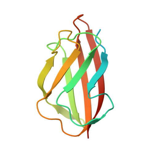
    The bacterial CopC family of proteins are periplasmic copper binding proteins that act in copper detoxification. These proteins contain Cu(I) and/or Cu(II) binding sites, with the family that binds Cu(II) only the most prevalent, based on sequence analyses. Here we present three crystal structures of the CopC protein from Pseudomonas fluorescens (Pf-CopC) that include the wild type protein bound to Cu(II) and two variant proteins, where Cu(II) coordinating ligands were mutated, in Cu-free states. We show that the Cu(II) atom in Pf-CopC is coordinated by two His residues, an Asp residue and the N-terminus of the protein (therefore a 3N + O site). This coordination structure is consistent with all structurally characterized proteins from the CopC family to date. Structural and sequence analyses of the CopC family allow a relationship between protein sequence and the Cu(II) binding affinity of these proteins to be proposed.


  • Organizational Affiliation

    Department of Biochemistry and Genetics, La Trobe Institute for Molecular Science, La Trobe University, Melbourne 3083, Australia.


Macromolecules
Find similar proteins by:  (by identity cutoff)  |  3D Structure
Entity ID: 1
MoleculeChains Sequence LengthOrganismDetailsImage
CopC
A, B, C
122Pseudomonas fluorescensMutation(s): 0 
UniProt
Find proteins for C3JYL7 (Pseudomonas fluorescens (strain SBW25))
Explore C3JYL7 
Go to UniProtKB:  C3JYL7
Entity Groups  
Sequence Clusters30% Identity50% Identity70% Identity90% Identity95% Identity100% Identity
UniProt GroupC3JYL7
Sequence Annotations
Expand
  • Reference Sequence
Experimental Data & Validation

Experimental Data

  • Method: X-RAY DIFFRACTION
  • Resolution: 2.00 Å
  • R-Value Free: 0.260 
  • R-Value Work: 0.228 
  • R-Value Observed: 0.230 
  • Space Group: C 2 2 21
Unit Cell:
Length ( Å )Angle ( ˚ )
a = 77.73α = 90
b = 111.319β = 90
c = 82.222γ = 90
Software Package:
Software NamePurpose
HKL-2000data reduction
SCALEPACKdata scaling
PHASERphasing
REFMACrefinement

Structure Validation

View Full Validation Report

Currently 6NFQ does not have a validation slider image.


Entry History & Funding Information

Deposition Data

  • Released Date: 2019-04-24 
  • Deposition Author(s): Maher, M.J.

Funding OrganizationLocationGrant Number
Australian Research Council (ARC)AustraliaDP140102746
Australian Research Council (ARC)AustraliaDP130100728

Revision History  (Full details and data files)

  • Version 1.0: 2019-04-24
    Type: Initial release
  • Version 1.1: 2020-01-01
    Changes: Author supporting evidence
  • Version 1.2: 2023-10-11
    Changes: Data collection, Database references, Derived calculations, Refinement description