6NBP

Crystal Structure of a Sugar N-Formyltransferase from the Plant Pathogen Pantoea ananatis


Experimental Data Snapshot

  • Method: X-RAY DIFFRACTION
  • Resolution: 1.70 Å
  • R-Value Free: 0.226 
  • R-Value Work: 0.186 
  • R-Value Observed: 0.188 

wwPDB Validation   3D Report Full Report

Currently 6NBP does not have a validation slider image.


This is version 1.5 of the entry. See complete history


Literature

Investigation of a sugar N-formyltransferase from the plant pathogen Pantoea ananatis.

Hofmeister, D.L.Thoden, J.B.Holden, H.M.

(2019) Protein Sci 28: 707-716

  • DOI: https://doi.org/10.1002/pro.3577
  • Primary Citation of Related Structures:  
    6NBP

  • PubMed Abstract: 

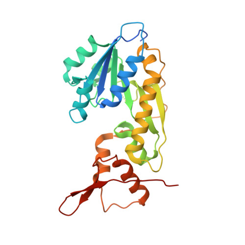
    Pantoea ananatis is a Gram-negative bacterium first recognized in 1928 as the causative agent of pineapple rot in the Philippines. Since then various strains of the organism have been implicated in the devastation of agriculturally important crops. Some strains, however, have been shown to function as non-pathogenic plant growth promoting organisms. To date, the factors that determine pathogenicity or lack thereof between the various strains are not well understood. All P. ananatis strains contain lipopolysaccharides, which differ with respect to the identities of their associated sugars. Given our research interest on the presence of the unusual sugar, 4-formamido-4,6-dideoxy-d-glucose, found on the lipopolysaccharides of Campylobacter jejuni and Francisella tularensis, we were curious as to whether other bacteria have the appropriate biosynthetic machinery to produce these unique carbohydrates. Four enzymes are typically required for their biosynthesis: a thymidylyltransferase, a 4,6-dehydratase, an aminotransferase, and an N-formyltransferase. Here, we report that the gene SAMN03097714_1080 from the P. ananatis strain NFR11 does, indeed, encode for an N-formyltransferase, hereafter referred to as PA1080c. Our kinetic analysis demonstrates that PA1080c displays classical Michaelis-Menten kinetics with dTDP-4-amino-4,6-dideoxy-d-glucose as the substrate and N 10 -formyltetrahydrofolate as the carbon source. In addition, the X-ray structure of PA1080c, determined to 1.7 Å resolution, shows that the enzyme adopts the molecular architecture observed for other sugar N-formyltransferases. Analysis of the P. ananatis NFR11 genome suggests that the three other enzymes necessary for N-formylated sugar biosynthesis are also present. Intriguingly, those strains of P. ananatis that are non-pathogenic apparently do not contain these genes.


  • Organizational Affiliation

    Department of Biochemistry, University of Wisconsin, Madison, Wisconsin, 53706.


Macromolecules
Find similar proteins by:  (by identity cutoff)  |  3D Structure
Entity ID: 1
MoleculeChains Sequence LengthOrganismDetailsImage
N-formyltransferase263Pantoea ananatisMutation(s): 0 
Gene Names: SAMN03097714_1080
UniProt
Find proteins for A0A4P1LYI6 (Pantoea ananas)
Explore A0A4P1LYI6 
Go to UniProtKB:  A0A4P1LYI6
Entity Groups  
Sequence Clusters30% Identity50% Identity70% Identity90% Identity95% Identity100% Identity
UniProt GroupA0A4P1LYI6
Sequence Annotations
Expand
  • Reference Sequence
Small Molecules
Ligands 4 Unique
IDChains Name / Formula / InChI Key2D Diagram3D Interactions
0FX (Subject of Investigation/LOI)
Query on 0FX

Download Ideal Coordinates CCD File 
B [auth A]dTDP-4-amino-4,6-dideoxyglucose
C16 H27 N3 O14 P2
UIVJXHWSIFBBCY-LPGAPTBISA-N
FON
Query on FON

Download Ideal Coordinates CCD File 
C [auth A]N-{[4-({[(6R)-2-amino-5-formyl-4-oxo-1,4,5,6,7,8-hexahydropteridin-6-yl]methyl}amino)phenyl]carbonyl}-L-glutamic acid
C20 H23 N7 O7
VVIAGPKUTFNRDU-OLZOCXBDSA-N
PO4
Query on PO4

Download Ideal Coordinates CCD File 
E [auth A]PHOSPHATE ION
O4 P
NBIIXXVUZAFLBC-UHFFFAOYSA-K
NA
Query on NA

Download Ideal Coordinates CCD File 
D [auth A]SODIUM ION
Na
FKNQFGJONOIPTF-UHFFFAOYSA-N
Experimental Data & Validation

Experimental Data

  • Method: X-RAY DIFFRACTION
  • Resolution: 1.70 Å
  • R-Value Free: 0.226 
  • R-Value Work: 0.186 
  • R-Value Observed: 0.188 
  • Space Group: P 31 2 1
Unit Cell:
Length ( Å )Angle ( ˚ )
a = 75.928α = 90
b = 75.928β = 90
c = 87.591γ = 120
Software Package:
Software NamePurpose
REFMACrefinement
SAINTdata reduction
SADABSdata scaling
PHASERphasing

Structure Validation

View Full Validation Report

Currently 6NBP does not have a validation slider image.


Entry History & Funding Information

Deposition Data


Funding OrganizationLocationGrant Number
National Institutes of Health/National Institute of General Medical Sciences (NIH/NIGMS)United StatesGM115921

Revision History  (Full details and data files)

  • Version 1.0: 2018-12-19
    Type: Initial release
  • Version 1.1: 2019-02-06
    Changes: Data collection, Database references
  • Version 1.2: 2019-04-03
    Changes: Data collection, Database references
  • Version 1.3: 2019-04-17
    Changes: Author supporting evidence, Data collection
  • Version 1.4: 2020-01-01
    Changes: Author supporting evidence
  • Version 1.5: 2023-10-11
    Changes: Data collection, Database references, Derived calculations, Refinement description