6N7O

Crystal structure of GIL01 gp7


Experimental Data Snapshot

  • Method: X-RAY DIFFRACTION
  • Resolution: 1.70 Å
  • R-Value Free: 0.230 
  • R-Value Work: 0.193 
  • R-Value Observed: 0.195 

wwPDB Validation   3D Report Full Report


This is version 1.3 of the entry. See complete history


Literature

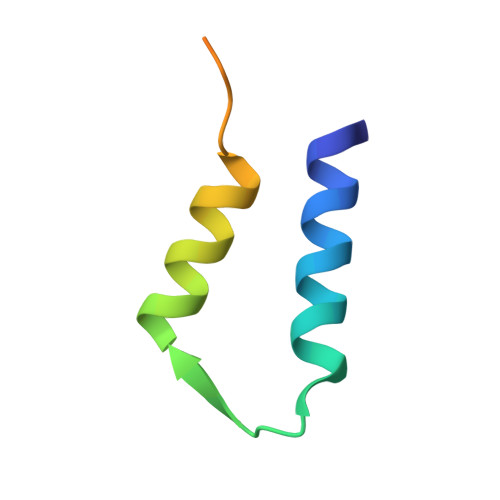
Structural Insights into Bacteriophage GIL01 gp7 Inhibition of Host LexA Repressor.

Caveney, N.A.Pavlin, A.Caballero, G.Bahun, M.Hodnik, V.de Castro, L.Fornelos, N.Butala, M.Strynadka, N.C.J.

(2019) Structure 27: 1094

  • DOI: https://doi.org/10.1016/j.str.2019.03.019
  • Primary Citation of Related Structures:  
    6N7O

  • PubMed Abstract: 

    Bacteria identify and respond to DNA damage using the SOS response. LexA, a central repressor in the response, has been implicated in the regulation of lysogeny in various temperate bacteriophages. During infection of Bacillus thuringiensis with GIL01 bacteriophage, LexA represses the SOS response and the phage lytic cycle by binding DNA, an interaction further stabilized upon binding of a viral protein, gp7. Here we report the crystallographic structure of phage-borne gp7 at 1.7-Å resolution, and characterize the 4:2 stoichiometry and potential interaction with LexA using surface plasmon resonance, static light scattering, and small-angle X-ray scattering. These data suggest that gp7 stabilizes LexA binding to operator DNA via coordination of the N- and C-terminal domains of LexA. Furthermore, we have found that gp7 can interact with LexA from Staphylococcus aureus, a significant human pathogen. Our results provide structural evidence as to how phage factors can directly associate with LexA to modulate the SOS response.


  • Organizational Affiliation

    Department of Biochemistry and Molecular Biology and the Centre for Blood Research, University of British Columbia, Vancouver, BC V6T 1Z3, Canada.


Macromolecules
Find similar proteins by:  (by identity cutoff)  |  3D Structure
Entity ID: 1
MoleculeChains Sequence LengthOrganismDetailsImage
GIL01 gp7A [auth B],
B [auth A]
52Bacillus phage pGIL01Mutation(s): 0 
UniProt
Find proteins for Q7WSG2 (Bacillus phage pGIL01)
Explore Q7WSG2 
Go to UniProtKB:  Q7WSG2
Entity Groups  
Sequence Clusters30% Identity50% Identity70% Identity90% Identity95% Identity100% Identity
UniProt GroupQ7WSG2
Sequence Annotations
Expand
  • Reference Sequence
Experimental Data & Validation

Experimental Data

  • Method: X-RAY DIFFRACTION
  • Resolution: 1.70 Å
  • R-Value Free: 0.230 
  • R-Value Work: 0.193 
  • R-Value Observed: 0.195 
  • Space Group: P 21 21 21
Unit Cell:
Length ( Å )Angle ( ˚ )
a = 39.69α = 90
b = 44.7β = 90
c = 46.3γ = 90
Software Package:
Software NamePurpose
PHENIXrefinement
XDSdata reduction
XSCALEdata scaling
PHASERphasing
PDB_EXTRACTdata extraction

Structure Validation

View Full Validation Report


Entry History & Funding Information

Deposition Data


Funding OrganizationLocationGrant Number
Canadian Institutes of Health Research (CIHR)Canada--

Revision History  (Full details and data files)

  • Version 1.0: 2019-05-15
    Type: Initial release
  • Version 1.1: 2019-07-17
    Changes: Data collection, Database references
  • Version 1.2: 2020-01-08
    Changes: Author supporting evidence
  • Version 1.3: 2024-03-13
    Changes: Data collection, Database references