


Crystal structure of a Tyrosine--tRNA ligase from Elizabethkingia anophelis
Edwards, T.E., Abendroth, J., Lorimer, D.D., Horanyi, P.S., Seattle Structural Genomics Center for Infectious Disease (SSGCID)To be published.
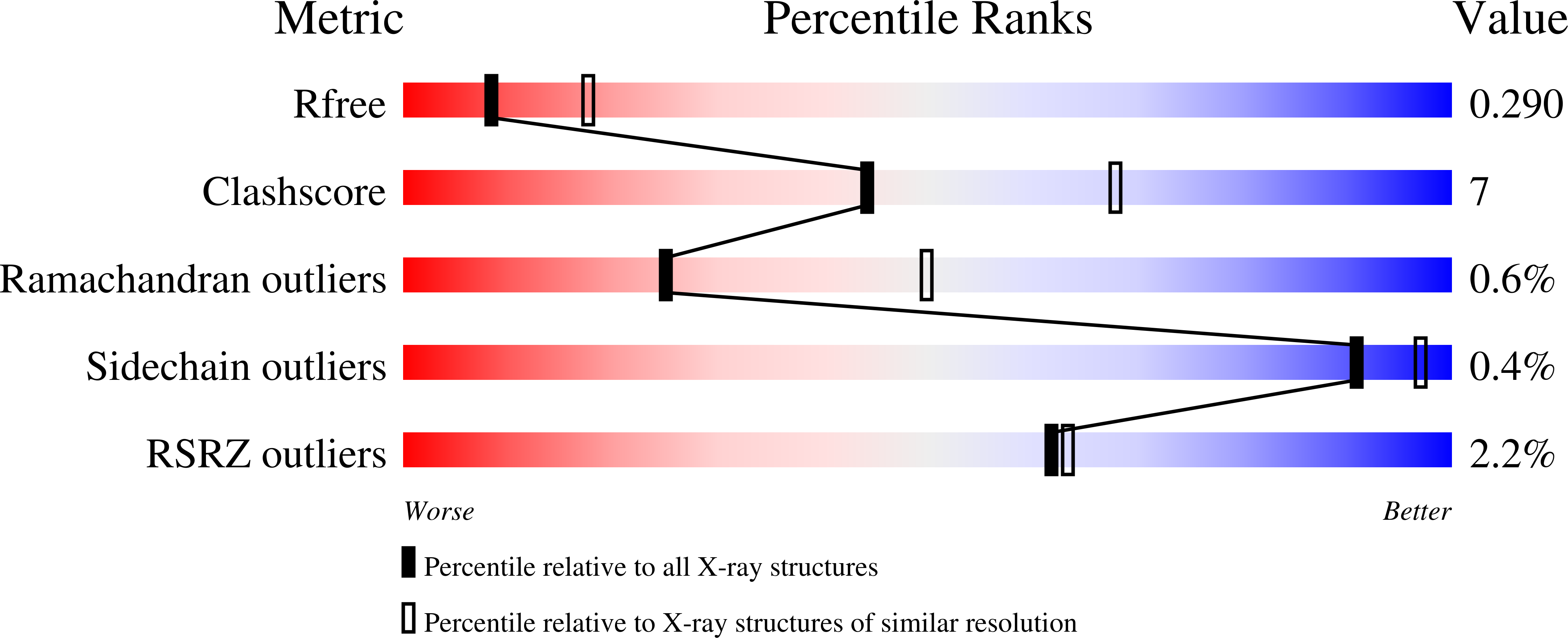
Experimental Data Snapshot
wwPDB Validation 3D Report Full Report
Entity ID: 1 | |||||
|---|---|---|---|---|---|
| Molecule | Chains | Sequence Length | Organism | Details | Image |
| Tyrosine--tRNA ligase | 480 | Elizabethkingia anophelis NUHP1 | Mutation(s): 0 Gene Names: tyrS, BD94_1246 EC: 6.1.1.1 | 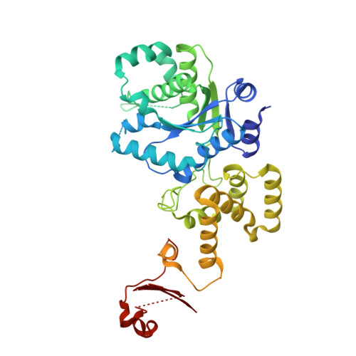 | |
UniProt | |||||
Find proteins for A0A077EBU6 (Elizabethkingia anophelis NUHP1) Explore A0A077EBU6 Go to UniProtKB: A0A077EBU6 | |||||
Entity Groups | |||||
| Sequence Clusters | 30% Identity50% Identity70% Identity90% Identity95% Identity100% Identity | ||||
| UniProt Group | A0A077EBU6 | ||||
Sequence AnnotationsExpand | |||||
| |||||
| Length ( Å ) | Angle ( ˚ ) |
|---|---|
| a = 197.29 | α = 90 |
| b = 37.56 | β = 112.4 |
| c = 81.76 | γ = 90 |
| Software Name | Purpose |
|---|---|
| XDS | data reduction |
| XSCALE | data scaling |
| MOLREP | phasing |
| PHENIX | refinement |
| PDB_EXTRACT | data extraction |
RCSB PDB (citation) is hosted by
RCSB PDB is a member of the

