6MXE

Crystal structure of human STING (G230A, H232R, R293Q) in complex with Compound 18


Experimental Data Snapshot

  • Method: X-RAY DIFFRACTION
  • Resolution: 2.47 Å
  • R-Value Free: 0.248 
  • R-Value Work: 0.196 
  • R-Value Observed: 0.199 

wwPDB Validation   3D Report Full Report


Ligand Structure Quality Assessment 


This is version 1.2 of the entry. See complete history


Literature

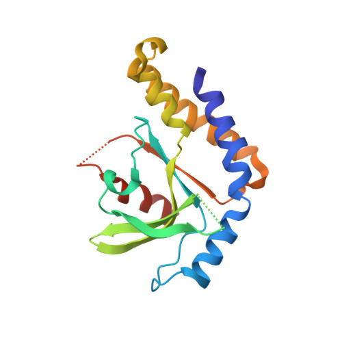
Discovery of a Novel cGAMP Competitive Ligand of the Inactive Form of STING.

Siu, T.Altman, M.D.Baltus, G.A.Childers, M.Ellis, J.M.Gunaydin, H.Hatch, H.Ho, T.Jewell, J.Lacey, B.M.Lesburg, C.A.Pan, B.S.Sauvagnat, B.Schroeder, G.K.Xu, S.

(2019) ACS Med Chem Lett 10: 92-97

  • DOI: https://doi.org/10.1021/acsmedchemlett.8b00466
  • Primary Citation of Related Structures:  
    6MX0, 6MX3, 6MXE

  • PubMed Abstract: 

    Drugging large protein pockets is a challenge due to the need for higher molecular weight ligands, which generally possess undesirable physicochemical properties. In this communication, we highlight a strategy leveraging small molecule active site dimers to inhibit the large symmetric binding pocket in the STING protein. By taking advantage of the 2:1 binding stoichiometry, maximal buried interaction with STING protein can be achieved while maintaining the ligand physicochemical properties necessary for oral exposure. This mode of binding requires unique considerations for potency optimization including simultaneous optimization of protein-ligand as well as ligand-ligand interactions. Successful implementation of this strategy led to the identification of 18 , which exhibits good oral exposure, slow binding kinetics, and functional inhibition of STING-mediated cytokine release.


  • Organizational Affiliation

    Departments of Chemistry, Immunology, Chemistry Modeling and Informatics, In Vitro Pharmacology, Target Protein Design and Structural Chemistry, Merck & Co., Inc., 33 Avenue Louis Pasteur, Boston, Massachusetts 02115, United States.


Macromolecules
Find similar proteins by:  (by identity cutoff)  |  3D Structure
Entity ID: 1
MoleculeChains Sequence LengthOrganismDetailsImage
Stimulator of interferon genes protein
A, B
188Homo sapiensMutation(s): 3 
Gene Names: TMEM173ERISMITASTING
UniProt & NIH Common Fund Data Resources
Find proteins for Q86WV6 (Homo sapiens)
Explore Q86WV6 
Go to UniProtKB:  Q86WV6
PHAROS:  Q86WV6
GTEx:  ENSG00000184584 
Entity Groups  
Sequence Clusters30% Identity50% Identity70% Identity90% Identity95% Identity100% Identity
UniProt GroupQ86WV6
Sequence Annotations
Expand
  • Reference Sequence
Small Molecules
Experimental Data & Validation

Experimental Data

  • Method: X-RAY DIFFRACTION
  • Resolution: 2.47 Å
  • R-Value Free: 0.248 
  • R-Value Work: 0.196 
  • R-Value Observed: 0.199 
  • Space Group: C 2 2 21
Unit Cell:
Length ( Å )Angle ( ˚ )
a = 69.85α = 90
b = 77.91β = 90
c = 124.35γ = 90
Software Package:
Software NamePurpose
BUSTERrefinement
Aimlessdata scaling
PDB_EXTRACTdata extraction

Structure Validation

View Full Validation Report


Ligand Structure Quality Assessment 


Entry History 

Deposition Data

Revision History  (Full details and data files)

  • Version 1.0: 2018-12-19
    Type: Initial release
  • Version 1.1: 2019-01-30
    Changes: Data collection, Database references
  • Version 1.2: 2024-03-13
    Changes: Data collection, Database references, Derived calculations