6MQU

PL5, synthetic transmembrane domain variant of human phospholamban


Experimental Data Snapshot

  • Method: X-RAY DIFFRACTION
  • Resolution: 3.17 Å
  • R-Value Free: 0.242 
  • R-Value Work: 0.228 
  • R-Value Observed: 0.229 

wwPDB Validation   3D Report Full Report


This is version 1.4 of the entry. See complete history


Literature

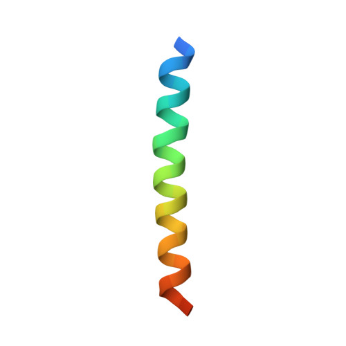
Packing of apolar side chains enables accurate design of highly stable membrane proteins.

Mravic, M.Thomaston, J.L.Tucker, M.Solomon, P.E.Liu, L.DeGrado, W.F.

(2019) Science 363: 1418-1423

  • DOI: https://doi.org/10.1126/science.aav7541
  • Primary Citation of Related Structures:  
    6MCT, 6MPW, 6MQ2, 6MQU

  • PubMed Abstract: 

    The features that stabilize the structures of membrane proteins remain poorly understood. Polar interactions contribute modestly, and the hydrophobic effect contributes little to the energetics of apolar side-chain packing in membranes. Disruption of steric packing can destabilize the native folds of membrane proteins, but is packing alone sufficient to drive folding in lipids? If so, then membrane proteins stabilized by this feature should be readily designed and structurally characterized-yet this has not been achieved. Through simulation of the natural protein phospholamban and redesign of variants, we define a steric packing code underlying its assembly. Synthetic membrane proteins designed using this code and stabilized entirely by apolar side chains conform to the intended fold. Although highly stable, the steric complementarity required for their folding is surprisingly stringent. Structural informatics shows that the designed packing motif recurs across the proteome, emphasizing a prominent role for precise apolar packing in membrane protein folding, stabilization, and evolution.


  • Organizational Affiliation

    Department of Pharmaceutical Chemistry, University of California, San Francisco, San Francisco, CA 94158, USA.


Macromolecules
Find similar proteins by:  (by identity cutoff)  |  3D Structure
Entity ID: 1
MoleculeChains Sequence LengthOrganismDetailsImage
PL5, designed TM pentamer
A, B, C, D, E
A, B, C, D, E, F, G, H, I, J
33synthetic constructMutation(s): 0 
Entity Groups  
Sequence Clusters30% Identity50% Identity70% Identity90% Identity95% Identity100% Identity
Sequence Annotations
Expand
  • Reference Sequence
Experimental Data & Validation

Experimental Data

  • Method: X-RAY DIFFRACTION
  • Resolution: 3.17 Å
  • R-Value Free: 0.242 
  • R-Value Work: 0.228 
  • R-Value Observed: 0.229 
  • Space Group: I 41
Unit Cell:
Length ( Å )Angle ( ˚ )
a = 124.23α = 90
b = 124.23β = 90
c = 51.94γ = 90
Software Package:
Software NamePurpose
MOSFLMdata reduction
Aimlessdata scaling
PHENIXphasing
PHENIXrefinement

Structure Validation

View Full Validation Report


Entry History & Funding Information

Deposition Data


Funding OrganizationLocationGrant Number
National Institutes of Health/National Institute of General Medical Sciences (NIH/NIGMS)United StatesR01 GM054616

Revision History  (Full details and data files)

  • Version 1.0: 2019-04-03
    Type: Initial release
  • Version 1.1: 2019-04-10
    Changes: Data collection, Database references
  • Version 1.2: 2019-04-17
    Changes: Data collection, Database references
  • Version 1.3: 2020-01-01
    Changes: Author supporting evidence
  • Version 1.4: 2024-03-13
    Changes: Data collection, Database references