6MLU

Cryo-EM structure of lipid droplet formation protein Seipin/BSCL2


Experimental Data Snapshot

  • Method: ELECTRON MICROSCOPY
  • Resolution: 4.00 Å
  • Aggregation State: PARTICLE 
  • Reconstruction Method: SINGLE PARTICLE 

wwPDB Validation   3D Report Full Report


This is version 1.4 of the entry. See complete history


Literature

Cryo-electron microscopy structure of the lipid droplet-formation protein seipin.

Sui, X.Arlt, H.Brock, K.P.Lai, Z.W.DiMaio, F.Marks, D.S.Liao, M.Farese Jr., R.V.Walther, T.C.

(2018) J Cell Biol 217: 4080-4091

  • DOI: https://doi.org/10.1083/jcb.201809067
  • Primary Citation of Related Structures:  
    6MLU

  • PubMed Abstract: 

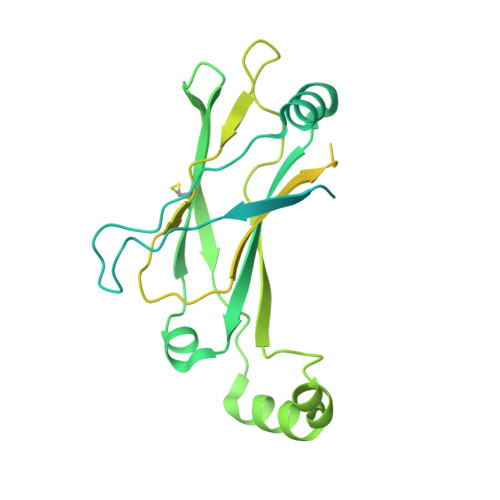
    Metabolic energy is stored in cells primarily as triacylglycerols in lipid droplets (LDs), and LD dysregulation leads to metabolic diseases. The formation of monolayer-bound LDs from the endoplasmic reticulum (ER) bilayer is poorly understood, but the ER protein seipin is essential to this process. In this study, we report a cryo-electron microscopy structure and functional characterization of Drosophila melanogaster seipin. The structure reveals a ring-shaped dodecamer with the luminal domain of each monomer resolved at ∼4.0 Å. Each luminal domain monomer exhibits two distinctive features: a hydrophobic helix (HH) positioned toward the ER bilayer and a β-sandwich domain with structural similarity to lipid-binding proteins. This structure and our functional testing in cells suggest a model in which seipin oligomers initially detect forming LDs in the ER via HHs and subsequently act as membrane anchors to enable lipid transfer and LD growth.


  • Organizational Affiliation

    Department of Cell Biology, Harvard Medical School, Boston, MA.


Macromolecules
Find similar proteins by:  (by identity cutoff)  |  3D Structure
Entity ID: 1
MoleculeChains Sequence LengthOrganismDetailsImage
Seipin
A, B
382Drosophila melanogasterMutation(s): 0 
Gene Names: SeipinCG9904
Membrane Entity: Yes 
UniProt
Find proteins for Q9V3X4 (Drosophila melanogaster)
Explore Q9V3X4 
Go to UniProtKB:  Q9V3X4
Entity Groups  
Sequence Clusters30% Identity50% Identity70% Identity90% Identity95% Identity100% Identity
UniProt GroupQ9V3X4
Sequence Annotations
Expand
  • Reference Sequence
Experimental Data & Validation

Experimental Data

  • Method: ELECTRON MICROSCOPY
  • Resolution: 4.00 Å
  • Aggregation State: PARTICLE 
  • Reconstruction Method: SINGLE PARTICLE 

Structure Validation

View Full Validation Report


Entry History & Funding Information

Deposition Data


Funding OrganizationLocationGrant Number
Howard Hughes Medical Institute (HHMI)United States--
National Institutes of Health/National Human Genome Research Institute (NIH/NHGRI)United States1R01GM123089
National Institutes of Health/National Human Genome Research Institute (NIH/NHGRI)United States1R01GM124348-01
National Institutes of Health/National Human Genome Research Institute (NIH/NHGRI)United StatesR01GM097194
American Heart AssociationUnited States18POST34030308
German Research Foundation (DFG)GermanyAR1164/1-1

Revision History  (Full details and data files)

  • Version 1.0: 2018-10-17
    Type: Initial release
  • Version 1.1: 2018-10-31
    Changes: Data collection, Database references
  • Version 1.2: 2018-12-12
    Changes: Data collection, Database references
  • Version 1.3: 2019-11-20
    Changes: Author supporting evidence
  • Version 1.4: 2019-12-18
    Changes: Other