


Crystal structure of carbon-nitrogen hydrolase from Helicobacter pylori G27
Abendroth, J., Lorimer, D.D., Horanyi, P.S., Edwards, T.E.To be published.
Experimental Data Snapshot
wwPDB Validation 3D Report Full Report

Entity ID: 1 | |||||
|---|---|---|---|---|---|
| Molecule | Chains | Sequence Length | Organism | Details | Image |
| Carbon-nitrogen hydrolase | 300 | Helicobacter pylori G27 | Mutation(s): 0 Gene Names: HPG27_713 | 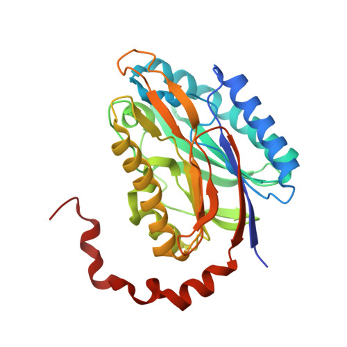 | |
UniProt | |||||
Find proteins for B5Z7B9 (Helicobacter pylori (strain G27)) Explore B5Z7B9 Go to UniProtKB: B5Z7B9 | |||||
Entity Groups | |||||
| Sequence Clusters | 30% Identity50% Identity70% Identity90% Identity95% Identity100% Identity | ||||
| UniProt Group | B5Z7B9 | ||||
Sequence AnnotationsExpand | |||||
| |||||
| Ligands 1 Unique | |||||
|---|---|---|---|---|---|
| ID | Chains | Name / Formula / InChI Key | 2D Diagram | 3D Interactions | |
| SO4 Query on SO4 | E [auth A] | SULFATE ION O4 S QAOWNCQODCNURD-UHFFFAOYSA-L |  | ||
| Length ( Å ) | Angle ( ˚ ) |
|---|---|
| a = 137.58 | α = 90 |
| b = 91.38 | β = 90 |
| c = 95.04 | γ = 90 |
| Software Name | Purpose |
|---|---|
| XDS | data reduction |
| XSCALE | data scaling |
| PHENIX | refinement |
| PDB_EXTRACT | data extraction |
| PHASER | phasing |
| PARROT | phasing |
| Coot | model building |
| ARP/wARP | model building |
RCSB PDB (citation) is hosted by
RCSB PDB is a member of the

