6MFV

Crystal structure of the Signal Transduction ATPase with Numerous Domains (STAND) protein with a tetratricopeptide repeat sensor PH0952 from Pyrococcus horikoshii


Experimental Data Snapshot

  • Method: X-RAY DIFFRACTION
  • Resolution: 3.40 Å
  • R-Value Free: 0.272 
  • R-Value Work: 0.224 
  • R-Value Observed: 0.226 

wwPDB Validation   3D Report Full Report


Ligand Structure Quality Assessment 


This is version 1.5 of the entry. See complete history


Literature

Double autoinhibition mechanism of signal transduction ATPases with numerous domains (STAND) with a tetratricopeptide repeat sensor.

Lisa, M.N.Cvirkaite-Krupovic, V.Richet, E.Andre-Leroux, G.Alzari, P.M.Haouz, A.Danot, O.

(2019) Nucleic Acids Res 47: 3795-3810

  • DOI: https://doi.org/10.1093/nar/gkz112
  • Primary Citation of Related Structures:  
    6MFV

  • PubMed Abstract: 

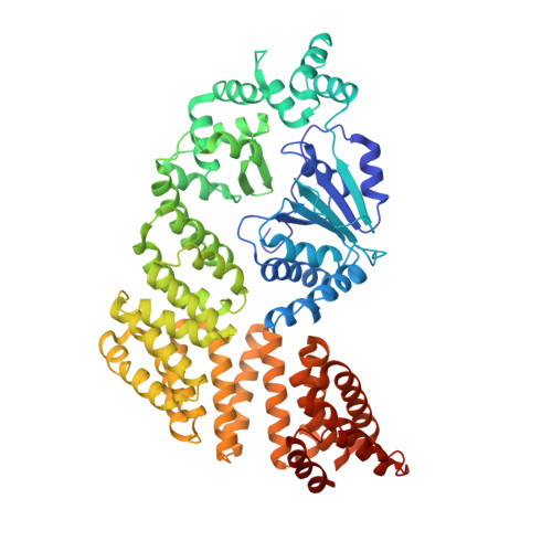
    Upon triggering by their inducer, signal transduction ATPases with numerous domains (STANDs), initially in monomeric resting forms, multimerize into large hubs that activate target macromolecules. This process requires conversion of the STAND conserved core (the NOD) from a closed form encasing an ADP molecule to an ATP-bound open form prone to multimerize. In the absence of inducer, autoinhibitory interactions maintain the NOD closed. In particular, in resting STAND proteins with an LRR- or WD40-type sensor domain, the latter establishes interactions with the NOD that are disrupted in the multimerization-competent forms. Here, we solved the first crystal structure of a STAND with a tetratricopeptide repeat sensor domain, PH0952 from Pyrococcus horikoshii, revealing analogous NOD-sensor contacts. We use this structural information to experimentally demonstrate that similar interactions also exist in a PH0952 homolog, the MalT STAND archetype, and actually contribute to the MalT autoinhibition in vitro and in vivo. We propose that STAND activation occurs by stepwise release of autoinhibitory contacts coupled to the unmasking of inducer-binding determinants. The MalT example suggests that STAND weak autoinhibitory interactions could assist the binding of inhibitory proteins by placing in register inhibitor recognition elements born by two domains.


  • Organizational Affiliation

    Unité de Microbiologie Structurale, Institut Pasteur, CNRS UMR 3528 & Université Paris Diderot, 75724 Paris Cedex 15, France.


Macromolecules
Find similar proteins by:  (by identity cutoff)  |  3D Structure
Entity ID: 1
MoleculeChains Sequence LengthOrganismDetailsImage
tetratricopeptide repeat sensor PH0952
A, B, C, D
653Pyrococcus horikoshii OT3Mutation(s): 0 
Gene Names: PH0952
UniProt
Find proteins for O58663 (Pyrococcus horikoshii (strain ATCC 700860 / DSM 12428 / JCM 9974 / NBRC 100139 / OT-3))
Explore O58663 
Go to UniProtKB:  O58663
Entity Groups  
Sequence Clusters30% Identity50% Identity70% Identity90% Identity95% Identity100% Identity
UniProt GroupO58663
Sequence Annotations
Expand
  • Reference Sequence
Experimental Data & Validation

Experimental Data

  • Method: X-RAY DIFFRACTION
  • Resolution: 3.40 Å
  • R-Value Free: 0.272 
  • R-Value Work: 0.224 
  • R-Value Observed: 0.226 
  • Space Group: P 65
Unit Cell:
Length ( Å )Angle ( ˚ )
a = 96.126α = 90
b = 96.126β = 90
c = 584.259γ = 120
Software Package:
Software NamePurpose
PHENIXrefinement
XDSdata reduction
Aimlessdata scaling
PHASERphasing

Structure Validation

View Full Validation Report


Ligand Structure Quality Assessment 


Entry History & Funding Information

Deposition Data


Funding OrganizationLocationGrant Number
French National Research AgencyFranceANR-08-BLAN-0204-01

Revision History  (Full details and data files)

  • Version 1.0: 2019-02-20
    Type: Initial release
  • Version 1.1: 2019-02-27
    Changes: Data collection, Database references
  • Version 1.2: 2019-03-06
    Changes: Data collection, Database references
  • Version 1.3: 2019-04-24
    Changes: Data collection, Database references
  • Version 1.4: 2024-03-13
    Changes: Data collection, Database references
  • Version 1.5: 2024-04-03
    Changes: Refinement description