


Crystal structure of PprA from Deinococcus radiodurans
Szabla, R., Czerwinski, M., Junop, M.S.To be published.
Experimental Data Snapshot
wwPDB Validation 3D Report Full Report

Entity ID: 1 | |||||
|---|---|---|---|---|---|
| Molecule | Chains | Sequence Length | Organism | Details | Image |
| DNA repair protein PprA | 308 | Deinococcus deserti VCD115 | Mutation(s): 2 Gene Names: pprA, Deide_2p01380 | 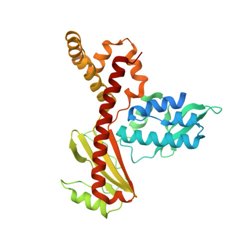 | |
UniProt | |||||
Find proteins for C1D318 (Deinococcus deserti (strain DSM 17065 / CIP 109153 / LMG 22923 / VCD115)) Explore C1D318 Go to UniProtKB: C1D318 | |||||
Entity Groups | |||||
| Sequence Clusters | 30% Identity50% Identity70% Identity90% Identity95% Identity100% Identity | ||||
| UniProt Group | C1D318 | ||||
Sequence AnnotationsExpand | |||||
| |||||
| Length ( Å ) | Angle ( ˚ ) |
|---|---|
| a = 54.417 | α = 90 |
| b = 80.985 | β = 90 |
| c = 148.868 | γ = 90 |
| Software Name | Purpose |
|---|---|
| PHENIX | refinement |
| CrysalisPro | data reduction |
| StructureStudio | data collection |
| PHASER | phasing |
| Aimless | data scaling |
| Funding Organization | Location | Grant Number |
|---|---|---|
| Natural Sciences and Engineering Research Council (NSERC, Canada) | Canada | 2008R00075 |
RCSB PDB (citation) is hosted by
RCSB PDB is a member of the

