6M9K

Crystal structure of lambda exonuclease in complex with the Red beta C-terminal domain


Experimental Data Snapshot

  • Method: X-RAY DIFFRACTION
  • Resolution: 2.30 Å
  • R-Value Free: 0.254 
  • R-Value Work: 0.195 
  • R-Value Observed: 0.198 

wwPDB Validation   3D Report Full Report


This is version 1.4 of the entry. See complete history


Literature

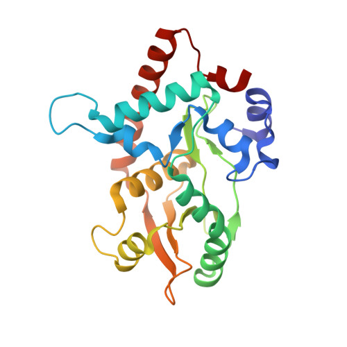
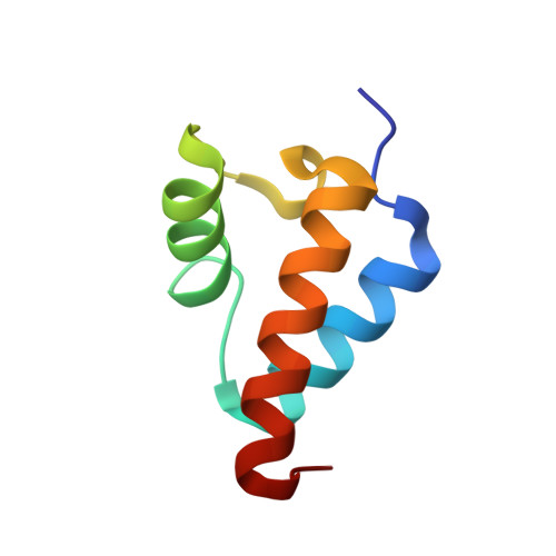
Crystal structure of the Red beta C-terminal domain in complex with lambda Exonuclease reveals an unexpected homology with lambda Orf and an interaction with Escherichia coli single stranded DNA binding protein.

Caldwell, B.J.Zakharova, E.Filsinger, G.T.Wannier, T.M.Hempfling, J.P.Chun-Der, L.Pei, D.Church, G.M.Bell, C.E.

(2019) Nucleic Acids Res 47: 1950-1963

  • DOI: https://doi.org/10.1093/nar/gky1309
  • Primary Citation of Related Structures:  
    6M9K

  • PubMed Abstract: 

    Bacteriophage λ encodes a DNA recombination system that includes a 5'-3' exonuclease (λ Exo) and a single strand annealing protein (Redβ). The two proteins form a complex that is thought to mediate loading of Redβ directly onto the single-stranded 3'-overhang generated by λ Exo. Here, we present a 2.3 Å crystal structure of the λ Exo trimer bound to three copies of the Redβ C-terminal domain (CTD). Mutation of residues at the hydrophobic core of the interface disrupts complex formation in vitro and impairs recombination in vivo. The Redβ CTD forms a three-helix bundle with unexpected structural homology to phage λ Orf, a protein that binds to E. coli single-stranded DNA binding protein (SSB) to function as a recombination mediator. Based on this relationship, we found that Redβ binds to full-length SSB, and to a peptide corresponding to its nine C-terminal residues, in an interaction that requires the CTD. These results suggest a dual role of the CTD, first in binding to λ Exo to facilitate loading of Redβ directly onto the initial single-stranded DNA (ssDNA) at a 3'-overhang, and second in binding to SSB to facilitate annealing of the overhang to SSB-coated ssDNA at the replication fork.


  • Organizational Affiliation

    Ohio State Biochemistry Program, The Ohio State University, 484 West 12th Avenue, Columbus, OH 43210, USA.


Macromolecules
Find similar proteins by:  (by identity cutoff)  |  3D Structure
Entity ID: 1
MoleculeChains Sequence LengthOrganismDetailsImage
Exonuclease
A, B, C
226Lambdavirus lambdaMutation(s): 0 
Gene Names: exored-alpharedX
EC: 3.1.11.3
UniProt
Find proteins for P03697 (Escherichia phage lambda)
Explore P03697 
Go to UniProtKB:  P03697
Entity Groups  
Sequence Clusters30% Identity50% Identity70% Identity90% Identity95% Identity100% Identity
UniProt GroupP03697
Sequence Annotations
Expand
  • Reference Sequence
Find similar proteins by:  (by identity cutoff)  |  3D Structure
Entity ID: 2
MoleculeChains Sequence LengthOrganismDetailsImage
Recombination protein bet
D, E, F
67Lambdavirus lambdaMutation(s): 0 
Gene Names: betbetAred-betaredB
UniProt
Find proteins for P03698 (Escherichia phage lambda)
Explore P03698 
Go to UniProtKB:  P03698
Entity Groups  
Sequence Clusters30% Identity50% Identity70% Identity90% Identity95% Identity100% Identity
UniProt GroupP03698
Sequence Annotations
Expand
  • Reference Sequence
Small Molecules
Ligands 1 Unique
IDChains Name / Formula / InChI Key2D Diagram3D Interactions
SO4
Query on SO4

Download Ideal Coordinates CCD File 
G [auth A]
H [auth A]
I [auth A]
J [auth A]
K [auth A]
G [auth A],
H [auth A],
I [auth A],
J [auth A],
K [auth A],
L [auth B],
M [auth B],
N [auth B],
O [auth B],
P [auth C],
Q [auth C],
R [auth C],
S [auth C],
T [auth C],
U [auth C],
V [auth F]
SULFATE ION
O4 S
QAOWNCQODCNURD-UHFFFAOYSA-L
Experimental Data & Validation

Experimental Data

  • Method: X-RAY DIFFRACTION
  • Resolution: 2.30 Å
  • R-Value Free: 0.254 
  • R-Value Work: 0.195 
  • R-Value Observed: 0.198 
  • Space Group: P 31 2 1
Unit Cell:
Length ( Å )Angle ( ˚ )
a = 122.522α = 90
b = 122.522β = 90
c = 147.858γ = 120
Software Package:
Software NamePurpose
HKL-2000data scaling
REFMACrefinement
PDB_EXTRACTdata extraction
HKL-2000data reduction
MOLREPphasing

Structure Validation

View Full Validation Report


Entry History & Funding Information

Deposition Data


Funding OrganizationLocationGrant Number
National Science Foundation (NSF, United States)United StatesMCB-1616105

Revision History  (Full details and data files)

  • Version 1.0: 2019-01-02
    Type: Initial release
  • Version 1.1: 2019-01-23
    Changes: Data collection, Database references
  • Version 1.2: 2019-03-13
    Changes: Data collection, Database references
  • Version 1.3: 2019-11-27
    Changes: Author supporting evidence
  • Version 1.4: 2023-10-11
    Changes: Data collection, Database references, Refinement description