Structure and functional analysis of a bacterial adhesin sugar-binding domain.
Vance, T.D.R., Guo, S., Assaie-Ardakany, S., Conroy, B., Davies, P.L.(2019) PLoS One 14: e0220045-e0220045
- PubMed: 31335890
- DOI: https://doi.org/10.1371/journal.pone.0220045
- Primary Citation of Related Structures:
6M8M - PubMed Abstract:
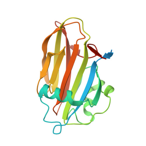
Bacterial adhesins attach their hosts to surfaces through one or more ligand-binding domains. In RTX adhesins, which are localized to the outer membrane of many Gram-negative bacteria via the type I secretion system, we see several examples of a putative sugar-binding domain. Here we have recombinantly expressed one such ~20-kDa domain from the ~340-kDa adhesin found in Marinobacter hydrocarbonoclasticus, an oil-degrading bacterium. The sugar-binding domain was purified from E. coli with a yield of 100 mg/L of culture. Circular dichroism analysis showed that the protein was rich in beta-structure, was moderately heat resistant, and required Ca2+ for proper folding. A crystal structure was obtained in Ca2+ at 1.2-Å resolution, which showed the presence of three Ca2+ ions, two of which were needed for structural integrity and one for binding sugars. Glucose was soaked into the crystal, where it bound to the sugar's two vicinal hydroxyl groups attached to the first and second (C1 and C2) carbons in the pyranose ring. This attraction to glucose caused the protein to bind certain polysaccharide-based column matrices and was used in a simple competitive binding assay to assess the relative affinity of sugars for the protein's ligand-binding site. Fucose, glucose and N-acetylglucosamine bound most tightly, and N-acetylgalactosamine hardly bound at all. Isothermal titration calorimetry was used to determine specific binding affinities, which lie in the 100-μM range. Glycan arrays were tested to expand the range of ligand sugars assayed, and showed that MhPA14 bound preferentially to branched polymers containing terminal sugars highlighted as strong binders in the competitive binding assay. Some of these binders have vicinal hydroxyl groups attached to the C3 and C4 carbons that are sterically equivalent to those presented by the C1 and C2 carbons of glucose.
Organizational Affiliation:
Department of Biomedical and Molecular Science, Queen's University, Kingston, Ontario, Canada.