6M77

GH31 alpha-N-acetylgalactosaminidase from Enterococcus faecalis in complex with N-acetylgalactosamine


Experimental Data Snapshot

  • Method: X-RAY DIFFRACTION
  • Resolution: 1.90 Å
  • R-Value Free: 0.261 
  • R-Value Work: 0.217 

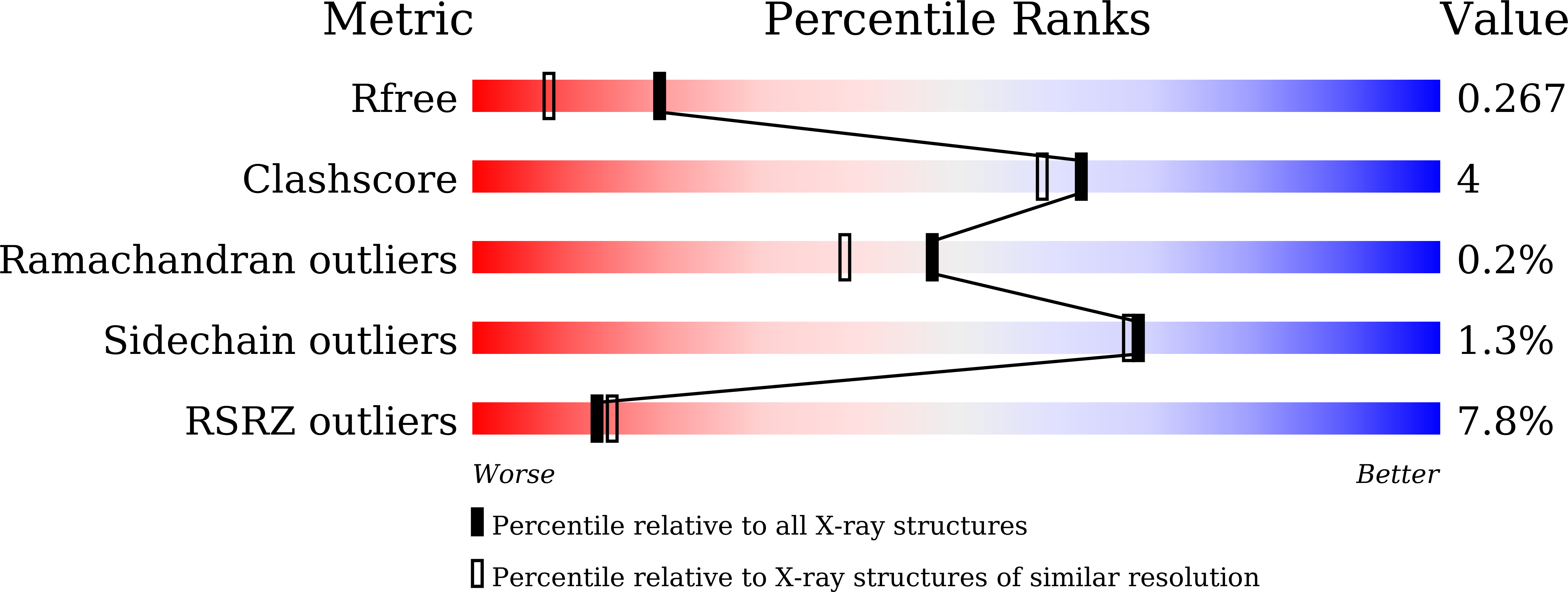
wwPDB Validation   3D Report Full Report


Ligand Structure Quality Assessment 


This is version 1.4 of the entry. See complete history


Literature

Crystal structure of the Enterococcus faecalis alpha-N-acetylgalactosaminidase, a member of the glycoside hydrolase family 31.

Miyazaki, T.Park, E.Y.

(2020) FEBS Lett 594: 2282-2293

  • DOI: https://doi.org/10.1002/1873-3468.13804
  • Primary Citation of Related Structures:  
    6M76, 6M77

  • PubMed Abstract: 

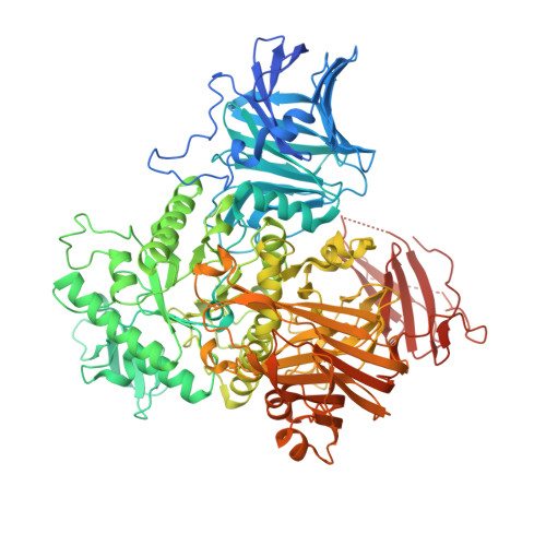
    Glycoside hydrolases catalyze the hydrolysis of glycosidic linkages in carbohydrates. The glycoside hydrolase family 31 (GH31) contains α-glucosidase, α-xylosidase, α-galactosidase, and α-transglycosylase. Recent work has expanded the diversity of substrate specificity of GH31 enzymes, and α-N-acetylgalactosaminidases (αGalNAcases) belonging to GH31 have been identified in human gut bacteria. Here, we determined the first crystal structure of a truncated form of GH31 αGalNAcase from the human gut bacterium Enterococcus faecalis. The enzyme has a similar fold to other reported GH31 enzymes and an additional fibronectin type 3-like domain. Additionally, the structure in complex with N-acetylgalactosamine reveals that conformations of the active site residues, including its catalytic nucleophile, change to recognize the ligand. Our structural analysis provides insight into the substrate recognition and catalytic mechanism of GH31 αGalNAcases.


  • Organizational Affiliation

    Green Chemistry Research Division, Research Institute of Green Science and Technology, Shizuoka University, Japan.


Macromolecules
Find similar proteins by:  (by identity cutoff)  |  3D Structure
Entity ID: 1
MoleculeChains Sequence LengthOrganismDetailsImage
LPXTG-motif cell wall anchor domain protein963Enterococcus faecalis ATCC 10100Mutation(s): 0 
UniProt
Find proteins for A0A125W693 (Enterococcus faecalis TX4248)
Explore A0A125W693 
Go to UniProtKB:  A0A125W693
Entity Groups  
Sequence Clusters30% Identity50% Identity70% Identity90% Identity95% Identity100% Identity
UniProt GroupA0A125W693
Sequence Annotations
Expand
  • Reference Sequence
Experimental Data & Validation

Experimental Data

  • Method: X-RAY DIFFRACTION
  • Resolution: 1.90 Å
  • R-Value Free: 0.261 
  • R-Value Work: 0.217 
  • Space Group: P 21 21 21
Unit Cell:
Length ( Å )Angle ( ˚ )
a = 82.596α = 90
b = 83.233β = 90
c = 147.787γ = 90
Software Package:
Software NamePurpose
REFMACrefinement
XDSdata reduction
XDSdata scaling
MOLREPphasing

Structure Validation

View Full Validation Report


Ligand Structure Quality Assessment 


Entry History & Funding Information

Deposition Data

  • Released Date: 2020-05-20 
  • Deposition Author(s): Miyazaki, T.

Funding OrganizationLocationGrant Number
Japan Society for the Promotion of Science (JSPS)Japan19K15748

Revision History  (Full details and data files)

  • Version 1.0: 2020-05-20
    Type: Initial release
  • Version 1.1: 2020-07-29
    Type: Remediation
    Reason: Carbohydrate remediation
    Changes: Data collection, Derived calculations, Structure summary
  • Version 1.2: 2020-08-12
    Changes: Database references, Structure summary
  • Version 1.3: 2020-09-16
    Changes: Database references, Source and taxonomy, Structure summary
  • Version 1.4: 2023-11-29
    Changes: Data collection, Database references, Derived calculations, Refinement description