6M55

Crystal structure of the E496A mutant of HsBglA in complex with 4-galactosyllactose


Experimental Data Snapshot

  • Method: X-RAY DIFFRACTION
  • Resolution: 3.00 Å
  • R-Value Free: 0.298 
  • R-Value Work: 0.265 
  • R-Value Observed: 0.266 

wwPDB Validation   3D Report Full Report


Ligand Structure Quality Assessment 


This is version 1.2 of the entry. See complete history


Literature

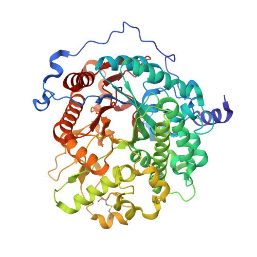
Crystal structure of a GH1 beta-glucosidase from Hamamotoa singularis.

Uehara, R.Iwamoto, R.Aoki, S.Yoshizawa, T.Takano, K.Matsumura, H.Tanaka, S.I.

(2020) Protein Sci 29: 2000-2008

  • DOI: https://doi.org/10.1002/pro.3916
  • Primary Citation of Related Structures:  
    6M4E, 6M4F, 6M55

  • PubMed Abstract: 

    A GH1 β-glucosidase from the fungus Hamamotoa singularis (HsBglA) has high transgalactosylation activity and efficiently converts lactose to galactooligosaccharides. Consequently, HsBglA is among the most widely used enzymes for industrial galactooligosaccharide production. Here, we present the first crystal structures of HsBglA with and without 4'-galactosyllactose, a tri-galactooligosaccharide, at 3.0 and 2.1 Å resolutions, respectively. These structures reveal details of the structural elements that define the catalytic activity and substrate binding of HsBglA, and provide a possible interpretation for its high catalytic potency for transgalactosylation reaction.


  • Organizational Affiliation

    Department of Biotechnology, College of Life Sciences, Ritsumeikan University, Shiga, Japan.


Macromolecules
Find similar proteins by:  (by identity cutoff)  |  3D Structure
Entity ID: 1
MoleculeChains Sequence LengthOrganismDetailsImage
Beta-galactosidase-like enzymeA,
B [auth D]
542Hamamotoa singularisMutation(s): 1 
Gene Names: bglA
UniProt
Find proteins for Q564N5 (Hamamotoa singularis)
Explore Q564N5 
Go to UniProtKB:  Q564N5
Entity Groups  
Sequence Clusters30% Identity50% Identity70% Identity90% Identity95% Identity100% Identity
UniProt GroupQ564N5
Sequence Annotations
Expand
  • Reference Sequence
Oligosaccharides

Help

Entity ID: 2
MoleculeChains Length2D Diagram Glycosylation3D Interactions
beta-D-galactopyranose-(1-4)-beta-D-galactopyranose-(1-4)-alpha-D-glucopyranoseC,
D [auth F]
3N/A
Glycosylation Resources
GlyTouCan:  G33559CW
GlyCosmos:  G33559CW
GlyGen:  G33559CW
Entity ID: 3
MoleculeChains Length2D Diagram Glycosylation3D Interactions
2-acetamido-2-deoxy-beta-D-glucopyranose-(1-4)-2-acetamido-2-deoxy-beta-D-glucopyranoseE [auth B],
F [auth E]
2N-Glycosylation
Glycosylation Resources
GlyTouCan:  G42666HT
GlyCosmos:  G42666HT
GlyGen:  G42666HT
Experimental Data & Validation

Experimental Data

  • Method: X-RAY DIFFRACTION
  • Resolution: 3.00 Å
  • R-Value Free: 0.298 
  • R-Value Work: 0.265 
  • R-Value Observed: 0.266 
  • Space Group: C 1 2 1
Unit Cell:
Length ( Å )Angle ( ˚ )
a = 184.686α = 90
b = 67.116β = 128.54
c = 117.585γ = 90
Software Package:
Software NamePurpose
PHENIXrefinement
PDB_EXTRACTdata extraction
XDSdata reduction
XSCALEdata scaling
PHASERphasing

Structure Validation

View Full Validation Report


Ligand Structure Quality Assessment 


Entry History & Funding Information

Deposition Data


Funding OrganizationLocationGrant Number
Ministry of Education, Culture, Sports, Science and Technology (Japan)Japan18K05445
Ministry of Education, Culture, Sports, Science and Technology (Japan)Japan19H05401

Revision History  (Full details and data files)

  • Version 1.0: 2020-09-02
    Type: Initial release
  • Version 1.1: 2020-12-16
    Changes: Database references
  • Version 1.2: 2023-11-29
    Changes: Data collection, Database references, Refinement description