6M3D

X-ray crystal structure of tandemly connected engrailed homeodomains (EHD) with R53A mutations and DNA complex


Experimental Data Snapshot

  • Method: X-RAY DIFFRACTION
  • Resolution: 1.60 Å
  • R-Value Free: 0.236 
  • R-Value Work: 0.188 
  • R-Value Observed: 0.191 

wwPDB Validation   3D Report Full Report


This is version 1.1 of the entry. See complete history


Literature

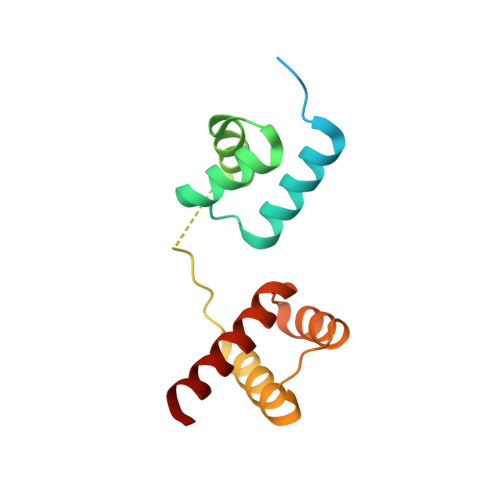
Structural basis for designing an array of engrailed homeodomains.

Sunami, T.Hirano, Y.Tamada, T.Kono, H.

(2020) Acta Crystallogr D Struct Biol 76: 824-833

  • DOI: https://doi.org/10.1107/S2059798320009237
  • Primary Citation of Related Structures:  
    6M3D

  • PubMed Abstract: 

    Small DNA-binding proteins that target desired sequences have the potential to act as a scaffold for molecular tools such as genome editing. In this study, an engrailed homeodomain (EHD) was chosen and it was evaluated whether it could be used as a molecular module that can connect to itself to recognize a longer target sequence. It was previously shown that two EHDs connected by a linker (EHD 2 ) recognize a target sequence twice as long as that recognized by a single EHD in cells only when Arg53 in each EHD in the tandem protein is mutated to alanine {(EHD[R53A]) 2 }. To investigate the recognition mechanism of (EHD[R53A]) 2 , the crystal structure of the (EHD[R53A]) 2 -DNA complex was determined at 1.6 Å resolution. The individual EHDs were found to adopt the typical homeodomain fold. Most importantly, the base-specific interactions in the major groove necessary for the affinity/specificity of wild-type EHD were preserved in (EHD[R53A]) 2 . Bacterial assays confirmed that the base-specific interactions are retained under cellular conditions. These observations indicate that the R53A mutation only causes a loss of the arginine-phosphate interaction at the protein-DNA interface, which reduces the DNA-binding affinity compared with the wild type. It is therefore concluded that (EHD[R53A]) 2 precisely recognizes tandem target sites within cells, enabling the individual EHDs to concurrently bind to the target sites with modest binding affinity. This suggests that modulation of the binding activity of each EHD is vital to construct a protein array that can precisely recognize a sequence with multiple target sites.


  • Organizational Affiliation

    Molecular Modeling and Simulation Group, National Institutes for Quantum and Radiological Science and Technology, 8-1-7 Umemidai, Kizugawa 619-0215, Japans.


Macromolecules

Find similar proteins by:  (by identity cutoff)  |  3D Structure
Entity ID: 3
MoleculeChains Sequence LengthOrganismDetailsImage
Segmentation polarity homeobox protein engrailed,Segmentation polarity homeobox protein engrailed148Drosophila melanogasterMutation(s): 4 
Gene Names: enCG9015
UniProt
Find proteins for P02836 (Drosophila melanogaster)
Explore P02836 
Go to UniProtKB:  P02836
Entity Groups  
Sequence Clusters30% Identity50% Identity70% Identity90% Identity95% Identity100% Identity
UniProt GroupP02836
Sequence Annotations
Expand
  • Reference Sequence

Find similar nucleic acids by:  Sequence   |   3D Structure  

Entity ID: 1
MoleculeChains LengthOrganismImage
DNA (5'-D(*TP*AP*AP*TP*CP*CP*TP*AP*AP*TP*CP*C)-3')12synthetic construct
Sequence Annotations
Expand
  • Reference Sequence

Find similar nucleic acids by:  Sequence   |   3D Structure  

Entity ID: 2
MoleculeChains LengthOrganismImage
DNA (5'-D(*GP*GP*AP*TP*TP*AP*GP*GP*AP*TP*TP*A)-3')12synthetic construct
Sequence Annotations
Expand
  • Reference Sequence
Small Molecules
Ligands 1 Unique
IDChains Name / Formula / InChI Key2D Diagram3D Interactions
NA
Query on NA

Download Ideal Coordinates CCD File 
D [auth A]SODIUM ION
Na
FKNQFGJONOIPTF-UHFFFAOYSA-N
Experimental Data & Validation

Experimental Data

  • Method: X-RAY DIFFRACTION
  • Resolution: 1.60 Å
  • R-Value Free: 0.236 
  • R-Value Work: 0.188 
  • R-Value Observed: 0.191 
  • Space Group: P 1 21 1
Unit Cell:
Length ( Å )Angle ( ˚ )
a = 39.798α = 90
b = 61.437β = 106.447
c = 39.976γ = 90
Software Package:
Software NamePurpose
PHENIXrefinement
XDSdata reduction
Aimlessdata scaling
PHASERphasing

Structure Validation

View Full Validation Report


Entry History & Funding Information

Deposition Data


Funding OrganizationLocationGrant Number
Japan Society for the Promotion of Science (JSPS)Japan26840054
Japan Society for the Promotion of Science (JSPS)Japan26330339
Japan Society for the Promotion of Science (JSPS)JapanJP18H05534

Revision History  (Full details and data files)

  • Version 1.0: 2020-09-16
    Type: Initial release
  • Version 1.1: 2023-11-29
    Changes: Data collection, Database references, Refinement description