6M1W

Structure of the 2-Aminoisobutyric acid Monooxygenase Hydroxylase


Experimental Data Snapshot

  • Method: X-RAY DIFFRACTION
  • Resolution: 2.75 Å
  • R-Value Free: 0.250 
  • R-Value Work: 0.190 
  • R-Value Observed: 0.193 

wwPDB Validation   3D Report Full Report


This is version 1.1 of the entry. See complete history

Re-refinement Note

A newer entry is available that reflects an alternative modeling of the original data: 6M2I


Literature

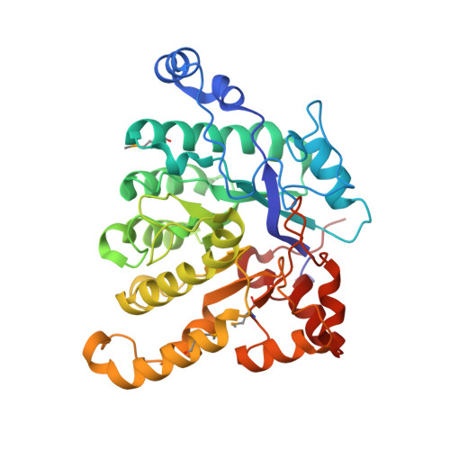
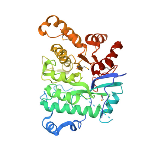
A three-component monooxygenase from Rhodococcus wratislaviensis may expand industrial applications of bacterial enzymes.

Hibi, M.Fukuda, D.Kenchu, C.Nojiri, M.Hara, R.Takeuchi, M.Aburaya, S.Aoki, W.Mizutani, K.Yasohara, Y.Ueda, M.Mikami, B.Takahashi, S.Ogawa, J.

(2021) Commun Biol 4: 16-16

  • DOI: https://doi.org/10.1038/s42003-020-01555-3
  • Primary Citation of Related Structures:  
    6M1W, 6M2I

  • PubMed Abstract: 

    The high-valent iron-oxo species formed in the non-heme diiron enzymes have high oxidative reactivity and catalyze difficult chemical reactions. Although the hydroxylation of inert methyl groups is an industrially promising reaction, utilizing non-heme diiron enzymes as such a biocatalyst has been difficult. Here we show a three-component monooxygenase system for the selective terminal hydroxylation of α-aminoisobutyric acid (Aib) into α-methyl-D-serine. It consists of the hydroxylase component, AibH1H2, and the electron transfer component. Aib hydroxylation is the initial step of Aib catabolism in Rhodococcus wratislaviensis C31-06, which has been fully elucidated through a proteome analysis. The crystal structure analysis revealed that AibH1H2 forms a heterotetramer of two amidohydrolase superfamily proteins, of which AibHm2 is a non-heme diiron protein and functions as a catalytic subunit. The Aib monooxygenase was demonstrated to be a promising biocatalyst that is suitable for bioprocesses in which the inert C-H bond in methyl groups need to be activated.


  • Organizational Affiliation

    Biotechnology Research Center and Department of Biotechnology, Toyama Prefectural University, 5180 Kurokawa, Imizu, Toyama, 939-0398, Japan. hibi@pu-toyama.ac.jp.


Macromolecules
Find similar proteins by:  (by identity cutoff)  |  3D Structure
Entity ID: 1
MoleculeChains Sequence LengthOrganismDetailsImage
Amidohydrolase354Rhodococcus wratislaviensisMutation(s): 0 
Gene Names: Rhow_000804
UniProt
Find proteins for A0A402C2V4 (Rhodococcus wratislaviensis)
Explore A0A402C2V4 
Go to UniProtKB:  A0A402C2V4
Entity Groups  
Sequence Clusters30% Identity50% Identity70% Identity90% Identity95% Identity100% Identity
UniProt GroupA0A402C2V4
Sequence Annotations
Expand
  • Reference Sequence
Find similar proteins by:  (by identity cutoff)  |  3D Structure
Entity ID: 2
MoleculeChains Sequence LengthOrganismDetailsImage
Amidohydrolase378Rhodococcus wratislaviensisMutation(s): 0 
Gene Names: Rhow_000803
UniProt
Find proteins for A0A402C2Q3 (Rhodococcus wratislaviensis)
Explore A0A402C2Q3 
Go to UniProtKB:  A0A402C2Q3
Entity Groups  
Sequence Clusters30% Identity50% Identity70% Identity90% Identity95% Identity100% Identity
UniProt GroupA0A402C2Q3
Sequence Annotations
Expand
  • Reference Sequence
Small Molecules
Ligands 5 Unique
IDChains Name / Formula / InChI Key2D Diagram3D Interactions
SO4
Query on SO4

Download Ideal Coordinates CCD File 
K [auth A]
L [auth A]
M [auth A]
N [auth A]
O [auth A]
K [auth A],
L [auth A],
M [auth A],
N [auth A],
O [auth A],
P [auth A],
U [auth B],
V [auth B]
SULFATE ION
O4 S
QAOWNCQODCNURD-UHFFFAOYSA-L
GOL
Query on GOL

Download Ideal Coordinates CCD File 
D [auth A]
E [auth A]
F [auth A]
G [auth A]
H [auth A]
D [auth A],
E [auth A],
F [auth A],
G [auth A],
H [auth A],
I [auth A],
J [auth A],
S [auth B],
T [auth B]
GLYCEROL
C3 H8 O3
PEDCQBHIVMGVHV-UHFFFAOYSA-N
ZN
Query on ZN

Download Ideal Coordinates CCD File 
C [auth A]ZINC ION
Zn
PTFCDOFLOPIGGS-UHFFFAOYSA-N
FE
Query on FE

Download Ideal Coordinates CCD File 
Q [auth B],
R [auth B]
FE (III) ION
Fe
VTLYFUHAOXGGBS-UHFFFAOYSA-N
CL
Query on CL

Download Ideal Coordinates CCD File 
W [auth B]CHLORIDE ION
Cl
VEXZGXHMUGYJMC-UHFFFAOYSA-M
Modified Residues  1 Unique
IDChains TypeFormula2D DiagramParent
MSE
Query on MSE
A
L-PEPTIDE LINKINGC5 H11 N O2 SeMET
Experimental Data & Validation

Experimental Data

  • Method: X-RAY DIFFRACTION
  • Resolution: 2.75 Å
  • R-Value Free: 0.250 
  • R-Value Work: 0.190 
  • R-Value Observed: 0.193 
  • Space Group: C 2 2 21
Unit Cell:
Length ( Å )Angle ( ˚ )
a = 125.49α = 90
b = 208.05β = 90
c = 90.66γ = 90
Software Package:
Software NamePurpose
PHENIXrefinement
XDSdata reduction
XDSdata scaling
PHENIXphasing

Structure Validation

View Full Validation Report


Entry History 

Deposition Data

Revision History  (Full details and data files)

  • Version 1.0: 2021-01-06
    Type: Initial release
  • Version 1.1: 2021-01-27
    Changes: Database references