6LZ1

Structure of S.pombe alpha-mannosidase Ams1


Experimental Data Snapshot

  • Method: ELECTRON MICROSCOPY
  • Resolution: 3.20 Å
  • Aggregation State: PARTICLE 
  • Reconstruction Method: SINGLE PARTICLE 

wwPDB Validation   3D Report Full Report


This is version 1.3 of the entry. See complete history


Literature

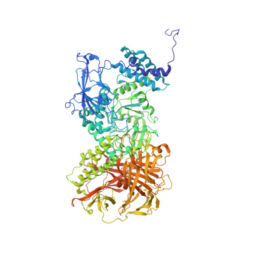
Cryo-EM structure of fission yeast tetrameric alpha-mannosidase Ams1.

Zhang, J.Wang, Y.Y.Du, L.L.Ye, K.

(2020) FEBS Open Bio 10: 2437-2451

  • DOI: https://doi.org/10.1002/2211-5463.12988
  • Primary Citation of Related Structures:  
    6LZ1

  • PubMed Abstract: 

    Fungal α-mannosidase Ams1 and its mammalian homolog MAN2C1 hydrolyze terminal α-linked mannoses in free oligosaccharides released from misfolded glycoproteins or lipid-linked oligosaccharide donors. Ams1 is transported by selective autophagy into vacuoles. Here, we determine the tetrameric structure of Ams1 from the fission yeast Schizosaccharomyces pombe at 3.2 Å resolution by cryo-electron microscopy. Distinct from a low resolution structure of S. cerevisiae Ams1, S. pombe Ams1 has a prominent N-terminal tail that mediates tetramerization and an extra β-sheet domain. Ams1 shares a conserved active site with other enzymes in glycoside hydrolase family 38, to which Ams1 belongs, but contains extra N-terminal domains involved in tetramerization. The atomic structure of Ams1 reported here will aid understanding of its enzymatic activity and transport mechanism.


  • Organizational Affiliation

    Key Laboratory of RNA Biology, CAS Center for Excellence in Biomacromolecules, Institute of Biophysics, Chinese Academy of Sciences, Beijing, China.


Macromolecules
Find similar proteins by:  (by identity cutoff)  |  3D Structure
Entity ID: 1
MoleculeChains Sequence LengthOrganismDetailsImage
Ams1
A, B, C, D
1,133Schizosaccharomyces pombe 972h-Mutation(s): 0 
Gene Names: ams1mns2SPAC513.05
EC: 3.2.1.24
UniProt
Find proteins for Q9UT61 (Schizosaccharomyces pombe (strain 972 / ATCC 24843))
Explore Q9UT61 
Go to UniProtKB:  Q9UT61
Entity Groups  
Sequence Clusters30% Identity50% Identity70% Identity90% Identity95% Identity100% Identity
UniProt GroupQ9UT61
Sequence Annotations
Expand
  • Reference Sequence
Experimental Data & Validation

Experimental Data

  • Method: ELECTRON MICROSCOPY
  • Resolution: 3.20 Å
  • Aggregation State: PARTICLE 
  • Reconstruction Method: SINGLE PARTICLE 
EM Software:
TaskSoftware PackageVersion
RECONSTRUCTIONRELION2.1
MODEL REFINEMENTPHENIX

Structure Validation

View Full Validation Report


Entry History & Funding Information

Deposition Data


Funding OrganizationLocationGrant Number
Ministry of Science and Technology (MoST, China)China2017YFA0504600
National Natural Science Foundation of China (NSFC)China91940302, 91540201, 31430024, 31325007
Chinese Academy of SciencesChinaXDB37010201

Revision History  (Full details and data files)

  • Version 1.0: 2020-09-09
    Type: Initial release
  • Version 1.1: 2020-10-28
    Changes: Database references
  • Version 1.2: 2021-03-10
    Changes: Database references
  • Version 1.3: 2024-03-27
    Changes: Data collection, Database references, Refinement description