6LXH

Staphylococcus aureus surface protein-sdrc


Experimental Data Snapshot

  • Method: X-RAY DIFFRACTION
  • Resolution: 2.07 Å
  • R-Value Free: 0.230 
  • R-Value Work: 0.170 
  • R-Value Observed: 0.173 

wwPDB Validation   3D Report Full Report


This is version 1.2 of the entry. See complete history


Literature

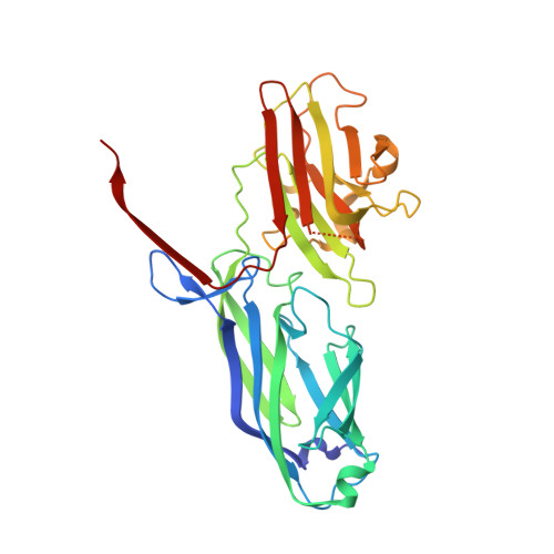
Structural Basis ofStaphylococcus aureusSurface Protein SdrC.

Pi, Y.Chen, W.Ji, Q.

(2020) Biochemistry 59: 1465-1469

  • DOI: https://doi.org/10.1021/acs.biochem.0c00124
  • Primary Citation of Related Structures:  
    6LXH, 6LXS

  • PubMed Abstract: 

    Staphylococcus aureus surface proteins play important roles in host tissue colonization, biofilm formation, and bacterial virulence and are thus essential for successful host infections. The surface protein SdrC from S. aureus induces bacterial biofilm formation via an intermolecular homophilic interaction of its N2 domains. However, the molecular mechanism of how the homophilic interaction is achieved is unknown. Here, we report two crystal structures of SdrC N2N3 domains, revealing two possible homophilic interaction mechanisms: Ca 2+ -mediated intermolecular metal chelation of N2 domains and intermolecular interaction of N2 and N3 domains. Given the unnecessary role of the N3 domain in the induction of biofilm formation, the N2 domain-mediated metal chelation mechanism is likely the mechanism that facilitates SdrC homophilic interaction. Mutation of key Ca 2+ -chelating residues differentially reduced the level of protein dimer formation, further supporting the key role of metal chelation in the N2 domain interaction. Together, these results reveal the possible mechanism of the homophilic interaction of SdrC N2 domains and pave the way for the rational development of new strategies against this mechanism.


  • Organizational Affiliation

    School of Physical Science and Technology, ShanghaiTech University, Shanghai 201210, China.


Macromolecules
Find similar proteins by:  (by identity cutoff)  |  3D Structure
Entity ID: 1
MoleculeChains Sequence LengthOrganismDetailsImage
Ser-Asp rich fibrinogen-binding, bone sialoprotein-binding protein
A, B, C
319Staphylococcus aureusMutation(s): 0 
Gene Names: sdrC
UniProt
Find proteins for Q2G0L5 (Staphylococcus aureus (strain NCTC 8325 / PS 47))
Explore Q2G0L5 
Go to UniProtKB:  Q2G0L5
Entity Groups  
Sequence Clusters30% Identity50% Identity70% Identity90% Identity95% Identity100% Identity
UniProt GroupQ2G0L5
Sequence Annotations
Expand
  • Reference Sequence
Experimental Data & Validation

Experimental Data

  • Method: X-RAY DIFFRACTION
  • Resolution: 2.07 Å
  • R-Value Free: 0.230 
  • R-Value Work: 0.170 
  • R-Value Observed: 0.173 
  • Space Group: P 21 21 21
Unit Cell:
Length ( Å )Angle ( ˚ )
a = 61.753α = 90
b = 120.333β = 90
c = 130.343γ = 90
Software Package:
Software NamePurpose
REFMACrefinement
HKL-2000data scaling
PDB_EXTRACTdata extraction
HKL-3000data reduction
HKL-3000phasing

Structure Validation

View Full Validation Report


Entry History 

Deposition Data

  • Released Date: 2020-04-22 
  • Deposition Author(s): Pi, Y., Ji, Q.

Revision History  (Full details and data files)

  • Version 1.0: 2020-04-22
    Type: Initial release
  • Version 1.1: 2020-05-06
    Changes: Database references
  • Version 1.2: 2024-03-27
    Changes: Data collection, Database references