6LW5

Crystal structure of the human formyl peptide receptor 2 in complex with WKYMVm


Experimental Data Snapshot

  • Method: X-RAY DIFFRACTION
  • Resolution: 2.80 Å
  • R-Value Free: 0.289 
  • R-Value Work: 0.258 

wwPDB Validation   3D Report Full Report


Ligand Structure Quality Assessment 


This is version 1.1 of the entry. See complete history


Literature

Structural basis of ligand binding modes at the human formyl peptide receptor 2.

Chen, T.Xiong, M.Zong, X.Ge, Y.Zhang, H.Wang, M.Won Han, G.Yi, C.Ma, L.Ye, R.D.Xu, Y.Zhao, Q.Wu, B.

(2020) Nat Commun 11: 1208-1208

  • DOI: https://doi.org/10.1038/s41467-020-15009-1
  • Primary Citation of Related Structures:  
    6LW5

  • PubMed Abstract: 

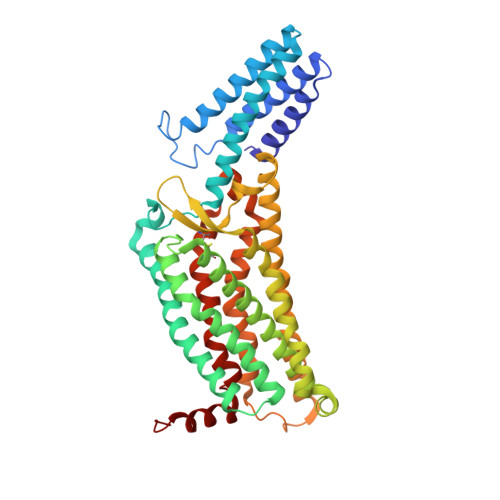
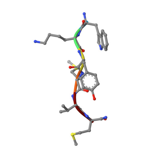
    The human formyl peptide receptor 2 (FPR2) plays a crucial role in host defense and inflammation, and has been considered as a drug target for chronic inflammatory diseases. A variety of peptides with different structures and origins have been characterized as FPR2 ligands. However, the ligand-binding modes of FPR2 remain elusive, thereby limiting the development of potential drugs. Here we report the crystal structure of FPR2 bound to the potent peptide agonist WKYMVm at 2.8 Å resolution. The structure adopts an active conformation and exhibits a deep ligand-binding pocket. Combined with mutagenesis, ligand binding and signaling studies, key interactions between the agonist and FPR2 that govern ligand recognition and receptor activation are identified. Furthermore, molecular docking and functional assays reveal key factors that may define binding affinity and agonist potency of formyl peptides. These findings deepen our understanding about ligand recognition and selectivity mechanisms of the formyl peptide receptor family.


  • Organizational Affiliation

    CAS Key Laboratory of Receptor Research, Shanghai Institute of Materia Medica, Chinese Academy of Sciences, 555 Zuchongzhi Road, 201203, Pudong, Shanghai, China.


Macromolecules
Find similar proteins by:  (by identity cutoff)  |  3D Structure
Entity ID: 1
MoleculeChains Sequence LengthOrganismDetailsImage
Soluble cytochrome b562,N-formyl peptide receptor 2427Escherichia coliHomo sapiens
This entity is chimeric
Mutation(s): 4 
Gene Names: cybCFPR2FPRH1FPRL1LXA4R
Membrane Entity: Yes 
UniProt & NIH Common Fund Data Resources
Find proteins for P0ABE7 (Escherichia coli)
Explore P0ABE7 
Go to UniProtKB:  P0ABE7
Find proteins for P25090 (Homo sapiens)
Explore P25090 
Go to UniProtKB:  P25090
PHAROS:  P25090
GTEx:  ENSG00000171049 
Entity Groups  
Sequence Clusters30% Identity50% Identity70% Identity90% Identity95% Identity100% Identity
UniProt GroupsP25090P0ABE7
Sequence Annotations
Expand
  • Reference Sequence

Find similar proteins by:  Sequence   |   3D Structure  

Entity ID: 2
MoleculeChains Sequence LengthOrganismDetailsImage
TRP-LYS-TYR-MET-VAL-QXV6synthetic constructMutation(s): 0 
Sequence Annotations
Expand
  • Reference Sequence
Experimental Data & Validation

Experimental Data

  • Method: X-RAY DIFFRACTION
  • Resolution: 2.80 Å
  • R-Value Free: 0.289 
  • R-Value Work: 0.258 
  • Space Group: P 32 2 1
Unit Cell:
Length ( Å )Angle ( ˚ )
a = 66.28α = 90
b = 66.28β = 90
c = 244.9γ = 120
Software Package:
Software NamePurpose
PHENIXrefinement
XDSdata reduction
XDSdata scaling
PHENIXphasing

Structure Validation

View Full Validation Report


Ligand Structure Quality Assessment 


Entry History & Funding Information

Deposition Data


Funding OrganizationLocationGrant Number
National Science Foundation (NSF, China)China31825010
Ministry of Science and Technology (MoST, China)China2018YFA0507000
National Science Foundation (NSF, China)China31730027
National Science Foundation (NSF, China)China81525024

Revision History  (Full details and data files)

  • Version 1.0: 2020-03-25
    Type: Initial release
  • Version 1.1: 2023-11-29
    Changes: Data collection, Database references, Refinement description