6LPI

Crystal Structure of AHAS holo-enzyme


Experimental Data Snapshot

  • Method: X-RAY DIFFRACTION
  • Resolution: 2.85 Å
  • R-Value Free: 0.243 
  • R-Value Work: 0.167 
  • R-Value Observed: 0.170 

wwPDB Validation   3D Report Full Report


Ligand Structure Quality Assessment 


This is version 1.1 of the entry. See complete history


Literature

Molecular architecture of the acetohydroxyacid synthase holoenzyme.

Zhang, Y.Li, Y.Liu, X.Sun, J.Li, X.Lin, J.Yang, X.Xi, Z.Shen, Y.

(2020) Biochem J 477: 2439-2449

  • DOI: https://doi.org/10.1042/BCJ20200292
  • Primary Citation of Related Structures:  
    6LPI

  • PubMed Abstract: 

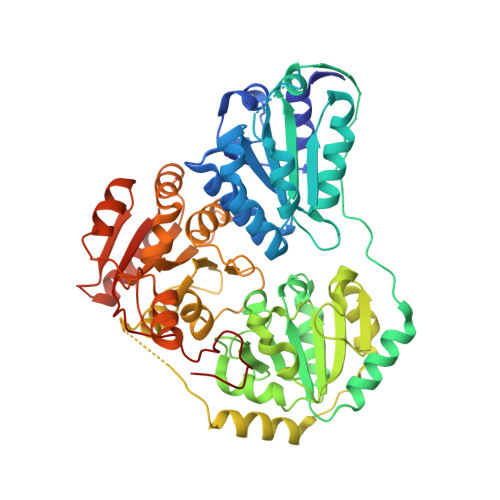
    The acetohydroxyacid synthase (AHAS) holoenzyme catalyzes the first step of branch-chain amino acid biosynthesis and is essential for plants and bacteria. It consists of a regulatory subunit (RSU) and a catalytic subunit (CSU). The allosteric mechanism of the AHAS holoenzyme has remained elusive for decades. Here, we determined the crystal structure of the AHAS holoenzyme, revealing the association between the RSU and CSU in an A2B2 mode. Structural analysis in combination with mutational studies demonstrated that the RSU dimer forms extensive interactions with the CSU dimer, in which a conserved salt bridge between R32 and D120 may act as a trigger to open the activation loop of the CSU, resulting in the activation of the CSU by the RSU. Our study reveals the activation mechanism of the AHAS holoenzyme.


  • Organizational Affiliation

    State Key Laboratory of Elemento-Organic Chemistry and College of Chemistry, Nankai University, Weijin 94, Tianjin 300071, China.


Macromolecules
Find similar proteins by:  (by identity cutoff)  |  3D Structure
Entity ID: 1
MoleculeChains Sequence LengthOrganismDetailsImage
Acetolactate synthase isozyme 1 small subunitA [auth E],
C [auth F],
E [auth G],
G [auth H]
127Escherichia coli K-12Mutation(s): 0 
Gene Names: ilvNb3670JW3645
EC: 2.2.1.6
UniProt
Find proteins for P0ADF8 (Escherichia coli (strain K12))
Explore P0ADF8 
Go to UniProtKB:  P0ADF8
Entity Groups  
Sequence Clusters30% Identity50% Identity70% Identity90% Identity95% Identity100% Identity
UniProt GroupP0ADF8
Sequence Annotations
Expand
  • Reference Sequence
Find similar proteins by:  (by identity cutoff)  |  3D Structure
Entity ID: 2
MoleculeChains Sequence LengthOrganismDetailsImage
Acetolactate synthase isozyme 1 large subunitB [auth A],
D [auth B],
F [auth C],
H [auth D]
562Escherichia coli K-12Mutation(s): 0 
Gene Names: ilvBb3671JW3646
EC: 2.2.1.6
UniProt
Find proteins for P08142 (Escherichia coli (strain K12))
Explore P08142 
Go to UniProtKB:  P08142
Entity Groups  
Sequence Clusters30% Identity50% Identity70% Identity90% Identity95% Identity100% Identity
UniProt GroupP08142
Sequence Annotations
Expand
  • Reference Sequence
Small Molecules
Ligands 3 Unique
IDChains Name / Formula / InChI Key2D Diagram3D Interactions
FAD (Subject of Investigation/LOI)
Query on FAD

Download Ideal Coordinates CCD File 
I [auth A],
L [auth B],
O [auth C],
R [auth D]
FLAVIN-ADENINE DINUCLEOTIDE
C27 H33 N9 O15 P2
VWWQXMAJTJZDQX-UYBVJOGSSA-N
TPP (Subject of Investigation/LOI)
Query on TPP

Download Ideal Coordinates CCD File 
J [auth A],
M [auth B],
P [auth C],
S [auth D]
THIAMINE DIPHOSPHATE
C12 H19 N4 O7 P2 S
AYEKOFBPNLCAJY-UHFFFAOYSA-O
MG (Subject of Investigation/LOI)
Query on MG

Download Ideal Coordinates CCD File 
K [auth A],
N [auth B],
Q [auth C],
T [auth D]
MAGNESIUM ION
Mg
JLVVSXFLKOJNIY-UHFFFAOYSA-N
Experimental Data & Validation

Experimental Data

  • Method: X-RAY DIFFRACTION
  • Resolution: 2.85 Å
  • R-Value Free: 0.243 
  • R-Value Work: 0.167 
  • R-Value Observed: 0.170 
  • Space Group: P 1 21 1
Unit Cell:
Length ( Å )Angle ( ˚ )
a = 111.173α = 90
b = 104.577β = 105.68
c = 118.535γ = 90
Software Package:
Software NamePurpose
PHENIXrefinement
PDB_EXTRACTdata extraction
HKL-2000data reduction
HKL-2000data scaling
PHASERphasing

Structure Validation

View Full Validation Report


Ligand Structure Quality Assessment 


Entry History 

Deposition Data

Revision History  (Full details and data files)

  • Version 1.0: 2020-11-18
    Type: Initial release
  • Version 1.1: 2023-11-29
    Changes: Data collection, Database references, Refinement description