6LHX

Crystal structure of ThsA


Experimental Data Snapshot

  • Method: X-RAY DIFFRACTION
  • Resolution: 2.50 Å
  • R-Value Free: 0.273 
  • R-Value Work: 0.242 
  • R-Value Observed: 0.242 

wwPDB Validation   3D Report Full Report


This is version 1.0 of the entry. See complete history


Literature

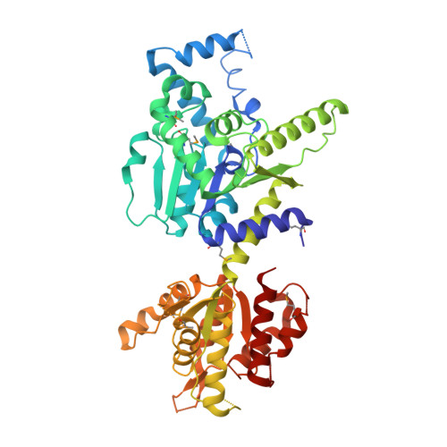
Structural and functional evidence of bacterial antiphage protection by Thoeris defense system via NAD+degradation.

Ka, D.Oh, H.Park, E.Kim, J.H.Bae, E.

(2020) Nat Commun 11: 2816-2816

  • DOI: https://doi.org/10.1038/s41467-020-16703-w
  • Primary Citation of Related Structures:  
    6LHX, 6LHY

  • PubMed Abstract: 

    The intense arms race between bacteria and phages has led to the development of diverse antiphage defense systems in bacteria. Unlike well-known restriction-modification and CRISPR-Cas systems, recently discovered systems are poorly characterized. One such system is the Thoeris defense system, which consists of two genes, thsA and thsB. Here, we report structural and functional analyses of ThsA and ThsB. ThsA exhibits robust NAD + cleavage activity and a two-domain architecture containing sirtuin-like and SLOG-like domains. Mutation analysis suggests that NAD + cleavage is linked to the antiphage function of Thoeris. ThsB exhibits a structural resemblance to TIR domain proteins such as nucleotide hydrolases and Toll-like receptors, but no enzymatic activity is detected in our in vitro assays. These results further our understanding of the molecular mechanism underlying the Thoeris defense system, highlighting a unique strategy for bacterial antiphage resistance via NAD + degradation.


  • Organizational Affiliation

    Department of Agricultural Biotechnology, Seoul National University, Seoul, 08826, Korea.


Macromolecules
Find similar proteins by:  (by identity cutoff)  |  3D Structure
Entity ID: 1
MoleculeChains Sequence LengthOrganismDetailsImage
ThsA
A, B, C, D
476Bacillus cereus MSX-D12Mutation(s): 0 
Gene Names: II9_05448
UniProt
Find proteins for J8G6Z1 (Bacillus cereus (strain MSX-D12))
Explore J8G6Z1 
Go to UniProtKB:  J8G6Z1
Entity Groups  
Sequence Clusters30% Identity50% Identity70% Identity90% Identity95% Identity100% Identity
UniProt GroupJ8G6Z1
Sequence Annotations
Expand
  • Reference Sequence
Small Molecules
Modified Residues  1 Unique
IDChains TypeFormula2D DiagramParent
MSE
Query on MSE
A, B, C, D
L-PEPTIDE LINKINGC5 H11 N O2 SeMET
Experimental Data & Validation

Experimental Data

  • Method: X-RAY DIFFRACTION
  • Resolution: 2.50 Å
  • R-Value Free: 0.273 
  • R-Value Work: 0.242 
  • R-Value Observed: 0.242 
  • Space Group: C 1 2 1
Unit Cell:
Length ( Å )Angle ( ˚ )
a = 180.424α = 90
b = 168.584β = 106.71
c = 93.614γ = 90
Software Package:
Software NamePurpose
PHENIXrefinement
HKL-2000data collection
HKL-2000data scaling
AutoSolphasing
PDB_EXTRACTdata extraction
PHENIXmodel building
BUCCANEERmodel building
HKL-2000data reduction

Structure Validation

View Full Validation Report


Entry History & Funding Information

Deposition Data


Funding OrganizationLocationGrant Number
National Research Foundation (NRF, Korea)Korea, Republic Of2019R1A2C1086298

Revision History  (Full details and data files)

  • Version 1.0: 2020-06-24
    Type: Initial release