6L7Q

Crystal structure of a hypothetical protein PYCH_01220 derived from Pyrococcus yayanosii


Experimental Data Snapshot

  • Method: X-RAY DIFFRACTION
  • Resolution: 1.16 Å
  • R-Value Free: 0.205 
  • R-Value Work: 0.192 
  • R-Value Observed: 0.192 

wwPDB Validation   3D Report Full Report


This is version 1.3 of the entry. See complete history


Literature

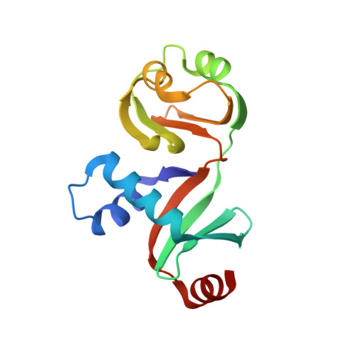
Crystal structure of PYCH_01220 from Pyrococcus yayanosii potentially involved in binding nucleic acid.

Noh, H.Jeon, J.H.Kim, Y.G.Oh, B.H.

(2021) Proteins 89: 468-472

  • DOI: https://doi.org/10.1002/prot.26029
  • Primary Citation of Related Structures:  
    6L7Q

  • PubMed Abstract: 

    We report the crystal structure of PYCH_01220, a hypothetical protein in Pyrococcus yayanosii CH1. This protein is composed of two domains, named Domain A and Domain B. While Domain B is not significantly homologous to known protein structures, Domain A is structurally analogous to the C-terminal ribonuclease domain of Escherichia coli colicin D. Domain A has a positively charged surface patch rendered by 13 basic residues, eight arginine or lysine residues of which are evolutionarily conserved. Electrophoretic mobility shift assays showed that PYCH_01220 binds to DNA, and charge-inversion mutations on this patch negatively affect the DNA binding, suggesting that the function of PYCH_01220 might involve nucleic acid-binding via the positively charged patch.


  • Organizational Affiliation

    Department of Biological Sciences, KAIST Institute for the Biocentury, Korea Advanced Institute of Science and Technology, Daejeon, Republic of Korea.


Macromolecules
Find similar proteins by:  (by identity cutoff)  |  3D Structure
Entity ID: 1
MoleculeChains Sequence LengthOrganismDetailsImage
hypothetical protein145Pyrococcus yayanosii CH1Mutation(s): 0 
Gene Names: PYCH_01220
UniProt
Find proteins for F8AFT0 (Pyrococcus yayanosii (strain CH1 / JCM 16557))
Explore F8AFT0 
Go to UniProtKB:  F8AFT0
Entity Groups  
Sequence Clusters30% Identity50% Identity70% Identity90% Identity95% Identity100% Identity
UniProt GroupF8AFT0
Sequence Annotations
Expand
  • Reference Sequence
Experimental Data & Validation

Experimental Data

  • Method: X-RAY DIFFRACTION
  • Resolution: 1.16 Å
  • R-Value Free: 0.205 
  • R-Value Work: 0.192 
  • R-Value Observed: 0.192 
  • Space Group: H 3
Unit Cell:
Length ( Å )Angle ( ˚ )
a = 86.278α = 90
b = 86.278β = 90
c = 62.438γ = 120
Software Package:
Software NamePurpose
PHENIXrefinement
HKL-2000data collection
HKL-2000data scaling
HKL-2000data reduction
PHENIXphasing
PDB_EXTRACTdata extraction

Structure Validation

View Full Validation Report


Entry History & Funding Information

Deposition Data


Funding OrganizationLocationGrant Number
National Research Foundation (NRF, Korea)Korea, Republic OfNRF-2018R1A2B3004764

Revision History  (Full details and data files)

  • Version 1.0: 2020-11-04
    Type: Initial release
  • Version 1.1: 2020-12-09
    Changes: Database references
  • Version 1.2: 2021-03-17
    Changes: Database references
  • Version 1.3: 2024-03-27
    Changes: Data collection, Database references