6L7D

Mycobacterium tuberculosis enolase mutant - S42A


Experimental Data Snapshot

  • Method: X-RAY DIFFRACTION
  • Resolution: 3.00 Å
  • R-Value Free: 0.243 
  • R-Value Work: 0.205 
  • R-Value Observed: 0.207 

wwPDB Validation   3D Report Full Report


Ligand Structure Quality Assessment 


This is version 3.2 of the entry. See complete history


Literature

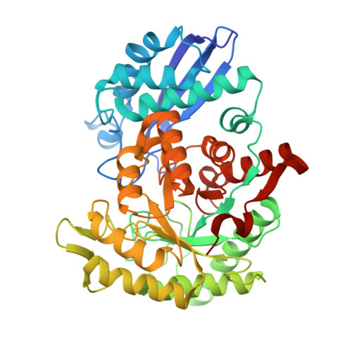
Structural snapshots of Mycobacterium tuberculosis enolase reveal dual mode of 2PG binding and its implication in enzyme catalysis.

Ahmad, M.Jha, B.Bose, S.Tiwari, S.Dwivedy, A.Kar, D.Pal, R.Mariadasse, R.Parish, T.Jeyakanthan, J.Vinothkumar, K.R.Biswal, B.K.

(2023) IUCrJ 10: 738-753

  • DOI: https://doi.org/10.1107/S2052252523008485
  • Primary Citation of Related Structures:  
    6L7D, 7CKP, 7CLK, 7CLL, 7DLR, 7E4F, 7E4X, 7E51

  • PubMed Abstract: 

    Enolase, a ubiquitous enzyme, catalyzes the reversible conversion of 2-phosphoglycerate (2PG) to phosphoenolpyruvate (PEP) in the glycolytic pathway of organisms of all three domains of life. The underlying mechanism of the 2PG to PEP conversion has been studied in great detail in previous work, however that of the reverse reaction remains to be explored. Here we present structural snapshots of Mycobacterium tuberculosis (Mtb) enolase in apo, PEP-bound and two 2PG-bound forms as it catalyzes the conversion of PEP to 2PG. The two 2PG-bound complex structures differed in the conformation of the bound product (2PG) viz the widely reported canonical conformation and a novel binding pose, which we refer to here as the alternate conformation. Notably, we observed two major differences compared with the forward reaction: the presence of Mg B is non-obligatory for the reaction and 2PG assumes an alternate conformation that is likely to facilitate its dissociation from the active site. Molecular dynamics studies and binding free energy calculations further substantiate that the alternate conformation of 2PG causes distortions in both metal ion coordination and hydrogen-bonding interactions, resulting in an increased flexibility of the active-site loops and aiding product release. Taken together, this study presents a probable mechanism involved in PEP to 2PG catalysis that is likely to be mediated by the conformational change of 2PG at the active site.


  • Organizational Affiliation

    Structural and Functional Biology Laboratory, National Institute of Immunology, Aruna Asaf Ali Marg, New Delhi 110067, India.


Macromolecules
Find similar proteins by:  (by identity cutoff)  |  3D Structure
Entity ID: 1
MoleculeChains Sequence LengthOrganismDetailsImage
Enolase436Mycobacterium tuberculosis H37RvMutation(s): 1 
Gene Names: enoRv1023MTCY10G2.26c
EC: 4.2.1.11
UniProt
Find proteins for P9WNL1 (Mycobacterium tuberculosis (strain ATCC 25618 / H37Rv))
Explore P9WNL1 
Go to UniProtKB:  P9WNL1
Entity Groups  
Sequence Clusters30% Identity50% Identity70% Identity90% Identity95% Identity100% Identity
UniProt GroupP9WNL1
Sequence Annotations
Expand
  • Reference Sequence
Experimental Data & Validation

Experimental Data

  • Method: X-RAY DIFFRACTION
  • Resolution: 3.00 Å
  • R-Value Free: 0.243 
  • R-Value Work: 0.205 
  • R-Value Observed: 0.207 
  • Space Group: I 4 2 2
Unit Cell:
Length ( Å )Angle ( ˚ )
a = 139.315α = 90
b = 139.315β = 90
c = 90.049γ = 90
Software Package:
Software NamePurpose
REFMACrefinement
PDB_EXTRACTdata extraction
HKL-2000data reduction
HKL-2000data scaling
PHASERphasing

Structure Validation

View Full Validation Report


Ligand Structure Quality Assessment 


Entry History & Funding Information

Deposition Data


Funding OrganizationLocationGrant Number
Department of Biotechnology (DBT, India)India--

Revision History  (Full details and data files)

  • Version 1.0: 2020-11-04
    Type: Initial release
  • Version 2.0: 2021-04-14
    Type: Coordinate replacement
    Reason: Ligand identity
    Changes: Advisory, Atomic model, Author supporting evidence, Data collection, Derived calculations, Non-polymer description, Other, Refinement description, Structure summary
  • Version 3.0: 2022-01-26
    Type: Coordinate replacement
    Reason: Ligand identity
    Changes: Advisory, Atomic model, Author supporting evidence, Data collection, Database references, Derived calculations, Non-polymer description, Other, Refinement description, Source and taxonomy, Structure summary
  • Version 3.1: 2023-11-08
    Changes: Data collection, Database references
  • Version 3.2: 2023-11-22
    Changes: Refinement description