


Heterochiral coupling in non-ribosomal peptide macrolactamization
Matsuda, K., Zhai, R., Mori, T., Kobayashi, M., Sano, A., Abe, I., Wakimoto, T.(2020) Nat Catal
Experimental Data Snapshot
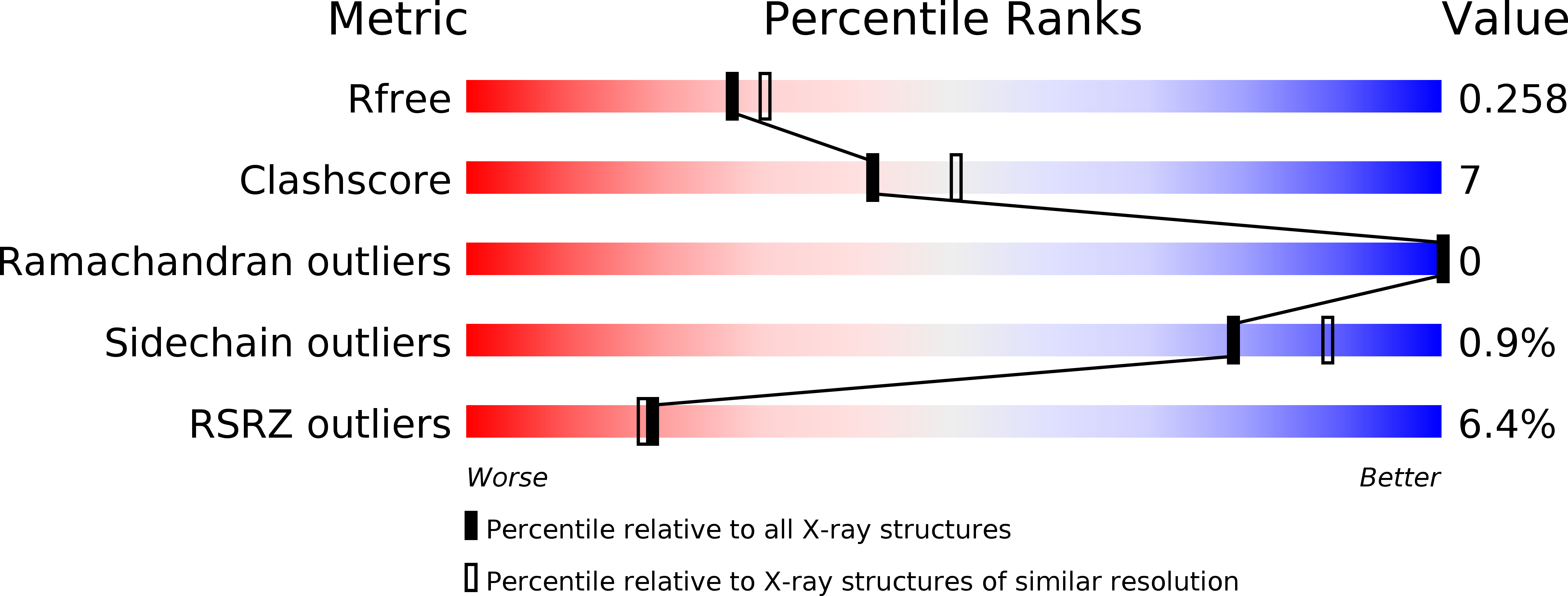
wwPDB Validation 3D Report Full Report
(2020) Nat Catal
Entity ID: 1 | |||||
|---|---|---|---|---|---|
| Molecule | Chains | Sequence Length | Organism | Details | Image |
| Alpha/beta hydrolase | 471 | Streptomyces albidoflavus | Mutation(s): 0 | 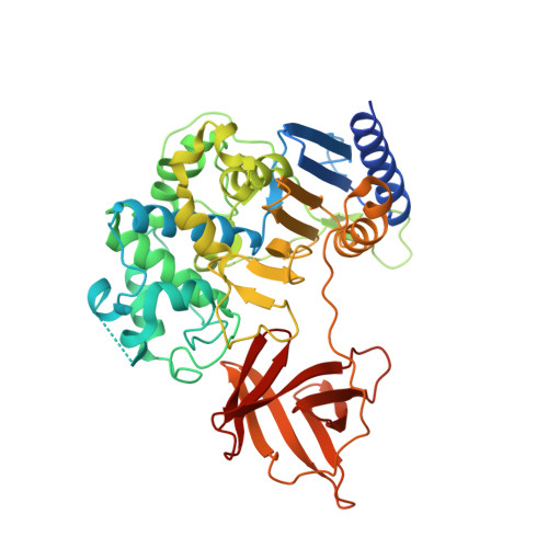 | |
UniProt | |||||
Find proteins for A0A679G4U8 (Streptomyces albidoflavus) Explore A0A679G4U8 Go to UniProtKB: A0A679G4U8 | |||||
Entity Groups | |||||
| Sequence Clusters | 30% Identity50% Identity70% Identity90% Identity95% Identity100% Identity | ||||
| UniProt Group | A0A679G4U8 | ||||
Sequence AnnotationsExpand | |||||
| |||||
| Ligands 3 Unique | |||||
|---|---|---|---|---|---|
| ID | Chains | Name / Formula / InChI Key | 2D Diagram | 3D Interactions | |
| MLI Query on MLI | D [auth A] | MALONATE ION C3 H2 O4 OFOBLEOULBTSOW-UHFFFAOYSA-L |  | ||
| SO4 Query on SO4 | C [auth A], E [auth B], G [auth B], H [auth B] | SULFATE ION O4 S QAOWNCQODCNURD-UHFFFAOYSA-L |  | ||
| YT3 Query on YT3 | F [auth B] | YTTRIUM (III) ION Y GRTBAGCGDOYUBE-UHFFFAOYSA-N |  | ||
| Length ( Å ) | Angle ( ˚ ) |
|---|---|
| a = 49.738 | α = 90 |
| b = 150.37 | β = 110.71 |
| c = 64.771 | γ = 90 |
| Software Name | Purpose |
|---|---|
| XSCALE | data scaling |
| PHENIX | refinement |
| PDB_EXTRACT | data extraction |
| XDS | data reduction |
| PHASER | phasing |
RCSB PDB (citation) is hosted by
RCSB PDB is a member of the

