6KOC

X-ray Structure of the proton-pumping cytochrome aa3-600 menaquinol oxidase from Bacillus subtilis complexed with 3-iodo-N-oxo-2-heptyl-4-hydroxyquinoline


Experimental Data Snapshot

  • Method: X-RAY DIFFRACTION
  • Resolution: 3.80 Å
  • R-Value Free: 0.369 
  • R-Value Work: 0.325 

wwPDB Validation   3D Report Full Report


Ligand Structure Quality Assessment 


This is version 1.3 of the entry. See complete history


Literature

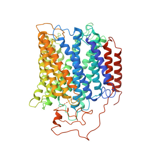
Structure of the cytochromeaa3-600 heme-copper menaquinol oxidase bound to inhibitor HQNO shows TM0 is part of the quinol binding site.

Xu, J.Ding, Z.Liu, B.Yi, S.M.Li, J.Zhang, Z.Liu, Y.Li, J.Liu, L.Zhou, A.Gennis, R.B.Zhu, J.

(2020) Proc Natl Acad Sci U S A 117: 872-876

  • DOI: https://doi.org/10.1073/pnas.1915013117
  • Primary Citation of Related Structures:  
    6KOB, 6KOC, 6KOE

  • PubMed Abstract: 

    Virtually all proton-pumping terminal respiratory oxygen reductases are members of the heme-copper oxidoreductase superfamily. Most of these enzymes use reduced cytochrome c as a source of electrons, but a group of enzymes have evolved to directly oxidize membrane-bound quinols, usually menaquinol or ubiquinol. All of the quinol oxidases have an additional transmembrane helix (TM0) in subunit I that is not present in the related cytochrome c oxidases. The current work reports the 3.6-Å-resolution X-ray structure of the cytochrome aa 3 -600 menaquinol oxidase from Bacillus subtilis containing 1 equivalent of menaquinone. The structure shows that TM0 forms part of a cleft to accommodate the menaquinol-7 substrate. Crystals which have been soaked with the quinol-analog inhibitor HQNO ( N -oxo-2-heptyl-4-hydroxyquinoline) or 3-iodo-HQNO reveal a single binding site where the inhibitor forms hydrogen bonds to amino acid residues shown previously by spectroscopic methods to interact with the semiquinone state of menaquinone, a catalytic intermediate.


  • Organizational Affiliation

    School of Medicine and Life Sciences, Nanjing University of Chinese Medicine, 210023 Nanjing, China.


Macromolecules
Find similar proteins by:  (by identity cutoff)  |  3D Structure
Entity ID: 1
MoleculeChains Sequence LengthOrganismDetailsImage
AA3-600 quinol oxidase subunit IA,
H [auth E]
649Bacillus subtilisMutation(s): 0 
Gene Names: qoxBB4122_4931B4417_2140ETA10_20065ETK61_21170ETL41_11350SC09_contig4orf01211
Membrane Entity: Yes 
UniProt
Find proteins for P34956 (Bacillus subtilis (strain 168))
Explore P34956 
Go to UniProtKB:  P34956
Entity Groups  
Sequence Clusters30% Identity50% Identity70% Identity90% Identity95% Identity100% Identity
UniProt GroupP34956
Sequence Annotations
Expand
  • Reference Sequence
Find similar proteins by:  (by identity cutoff)  |  3D Structure
Entity ID: 2
MoleculeChains Sequence LengthOrganismDetailsImage
Quinol oxidase subunit 2B,
G [auth F]
296Bacillus subtilisMutation(s): 0 
Gene Names: qoxAETA10_20070ETK61_21175ETL41_11355
EC: 1.10.3
Membrane Entity: Yes 
UniProt
Find proteins for P34957 (Bacillus subtilis (strain 168))
Explore P34957 
Go to UniProtKB:  P34957
Entity Groups  
Sequence Clusters30% Identity50% Identity70% Identity90% Identity95% Identity100% Identity
UniProt GroupP34957
Sequence Annotations
Expand
  • Reference Sequence
Find similar proteins by:  (by identity cutoff)  |  3D Structure
Entity ID: 3
MoleculeChains Sequence LengthOrganismDetailsImage
AA3-600 quinol oxidase subunit IIIIC,
E [auth G]
204Bacillus subtilisMutation(s): 0 
Gene Names: B4122_4930B4417_2139ETA10_20060ETK61_21165ETL41_11345SC09_contig4orf01209
Membrane Entity: Yes 
UniProt
Find proteins for P34958 (Bacillus subtilis (strain 168))
Explore P34958 
Go to UniProtKB:  P34958
Entity Groups  
Sequence Clusters30% Identity50% Identity70% Identity90% Identity95% Identity100% Identity
UniProt GroupP34958
Sequence Annotations
Expand
  • Reference Sequence
Find similar proteins by:  (by identity cutoff)  |  3D Structure
Entity ID: 4
MoleculeChains Sequence LengthOrganismDetailsImage
Quinol oxidase subunit 4,Quinol oxidase subunit 4D,
F [auth H]
123Bacillus subtilis subsp. subtilis str. 168Mutation(s): 0 
EC: 1.10.3
Membrane Entity: Yes 
UniProt
Find proteins for P34959 (Bacillus subtilis (strain 168))
Explore P34959 
Go to UniProtKB:  P34959
Entity Groups  
Sequence Clusters30% Identity50% Identity70% Identity90% Identity95% Identity100% Identity
UniProt GroupP34959
Sequence Annotations
Expand
  • Reference Sequence
Small Molecules
Ligands 3 Unique
IDChains Name / Formula / InChI Key2D Diagram3D Interactions
HEA
Query on HEA

Download Ideal Coordinates CCD File 
I [auth A],
J [auth A],
M [auth E],
N [auth E]
HEME-A
C49 H56 Fe N4 O6
ZGGYGTCPXNDTRV-PRYGPKJJSA-L
IHQ
Query on IHQ

Download Ideal Coordinates CCD File 
L [auth A],
P [auth E]
2-heptyl-3-iodanyl-1-oxidanyl-quinolin-4-one
C16 H20 I N O2
KWUZRGREQVAAQR-UHFFFAOYSA-N
CU
Query on CU

Download Ideal Coordinates CCD File 
K [auth A],
O [auth E]
COPPER (II) ION
Cu
JPVYNHNXODAKFH-UHFFFAOYSA-N
Experimental Data & Validation

Experimental Data

  • Method: X-RAY DIFFRACTION
  • Resolution: 3.80 Å
  • R-Value Free: 0.369 
  • R-Value Work: 0.325 
  • Space Group: P 1 21 1
Unit Cell:
Length ( Å )Angle ( ˚ )
a = 113.93α = 90
b = 162.2β = 109.76
c = 151.32γ = 90
Software Package:
Software NamePurpose
Aimlessdata scaling
PHENIXrefinement
PDB_EXTRACTdata extraction
iMOSFLMdata reduction
PHASERphasing

Structure Validation

View Full Validation Report


Ligand Structure Quality Assessment 


Entry History 

Deposition Data

Revision History  (Full details and data files)

  • Version 1.0: 2020-01-15
    Type: Initial release
  • Version 1.1: 2020-01-22
    Changes: Database references
  • Version 1.2: 2020-01-29
    Changes: Database references
  • Version 1.3: 2023-11-22
    Changes: Data collection, Database references, Refinement description