6KMM

Crystal Structure of HEPES bound Dye Decolorizing peroxidase from Bacillus subtilis


Experimental Data Snapshot

  • Method: X-RAY DIFFRACTION
  • Resolution: 1.93 Å
  • R-Value Free: 0.202 
  • R-Value Work: 0.161 
  • R-Value Observed: 0.163 

wwPDB Validation   3D Report Full Report


Ligand Structure Quality Assessment 


This is version 1.2 of the entry. See complete history


Literature

Characterization of dye-decolorizing peroxidase from Bacillus subtilis.

Dhankhar, P.Dalal, V.Mahto, J.K.Gurjar, B.R.Tomar, S.Sharma, A.K.Kumar, P.

(2020) Arch Biochem Biophys 693: 108590-108590

  • DOI: https://doi.org/10.1016/j.abb.2020.108590
  • Primary Citation of Related Structures:  
    6KMM, 6KMN

  • PubMed Abstract: 

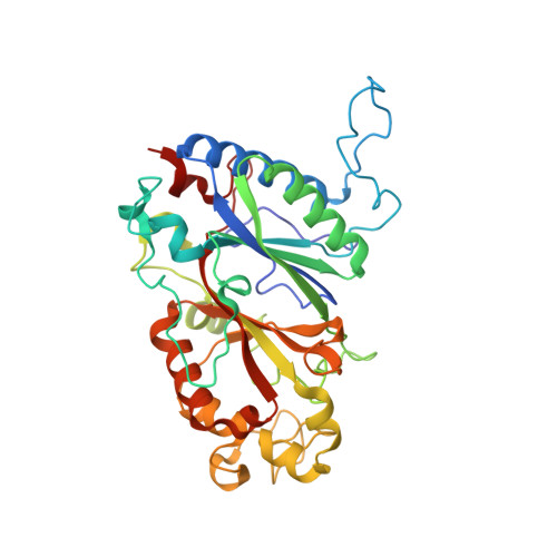
    The dye-decolorizing peroxidases (DyPs) belong to a unique heme peroxidase family for their biotechnological potential to detoxify synthetic dyes. In this work, we have biochemically and structurally characterized the dye-decolorizing peroxidase from Bacillus subtilis (BsDyP). The biochemical studies of BsDyP demonstrate that pH 4.0 is optimum for the oxidation of malachite green (MG) and methyl violet (MV). However, it oxidizes the MG with higher catalytic efficiency (k cat /K m  = 6.3 × 10 2 M -1 s -1 ), than MV (k cat /K m  = 5.0 × 10 2 M -1 s -1 ). While reactive black 5 (RB5) is oxidized at pH 3.0 with the catalytic efficiency of k cat /K m  = 3.6 × 10 2 M -1 s -1 . The calculated thermodynamic parameters by isothermal titration calorimetry (ITC) reveal the feasibility and spontaneity of dyes binding with BsDyP. Further, the crystal structures of a HEPES bound and unbound of BsDyP provide insight into the probable binding sites of the substrates. In BsDyP-HEPES bound structure, the HEPES-1 molecule is found in the heme cavity at the γ-edge, and another HEPES-2 molecule is bound ~16 Å away from the heme that is fenced by Ile231, Arg234, Ser235, Asp239, Glu334, and surface-exposed Tyr335 residues. Furthermore, the molecular docking, simulation, and MMPBSA studies support the binding of dyes at both the sites of BsDyP and produce lower-energy stable BsDyP-dyes complexes. Here, the BsDyP study allows the identification of its two potential binding sites and shows the oxidation of a variety of dyes. Structural and functional insight of BsDyP will facilitate its engineering for the improved decolorization of dyes.


  • Organizational Affiliation

    Department of Biotechnology, Indian Institute of Technology Roorkee, 247667, India.


Macromolecules
Find similar proteins by:  (by identity cutoff)  |  3D Structure
Entity ID: 1
MoleculeChains Sequence LengthOrganismDetailsImage
Deferrochelatase/peroxidase EfeB
A, B, C, D, E
A, B, C, D, E, F
363Bacillus subtilisMutation(s): 0 
Gene Names: efeBFC605_19685
UniProt
Find proteins for P39597 (Bacillus subtilis (strain 168))
Explore P39597 
Go to UniProtKB:  P39597
Entity Groups  
Sequence Clusters30% Identity50% Identity70% Identity90% Identity95% Identity100% Identity
UniProt GroupP39597
Sequence Annotations
Expand
  • Reference Sequence
Small Molecules
Ligands 8 Unique
IDChains Name / Formula / InChI Key2D Diagram3D Interactions
HEM (Subject of Investigation/LOI)
Query on HEM

Download Ideal Coordinates CCD File 
DA [auth E]
G [auth A]
HA [auth F]
M [auth B]
Q [auth C]
DA [auth E],
G [auth A],
HA [auth F],
M [auth B],
Q [auth C],
X [auth D]
PROTOPORPHYRIN IX CONTAINING FE
C34 H32 Fe N4 O4
KABFMIBPWCXCRK-RGGAHWMASA-L
EPE
Query on EPE

Download Ideal Coordinates CCD File 
AA [auth D],
I [auth A],
JA [auth F],
S [auth C],
Z [auth D]
4-(2-HYDROXYETHYL)-1-PIPERAZINE ETHANESULFONIC ACID
C8 H18 N2 O4 S
JKMHFZQWWAIEOD-UHFFFAOYSA-N
MPD
Query on MPD

Download Ideal Coordinates CCD File 
BA [auth D],
P [auth B],
V [auth C]
(4S)-2-METHYL-2,4-PENTANEDIOL
C6 H14 O2
SVTBMSDMJJWYQN-YFKPBYRVSA-N
PO4
Query on PO4

Download Ideal Coordinates CCD File 
U [auth C]PHOSPHATE ION
O4 P
NBIIXXVUZAFLBC-UHFFFAOYSA-K
GOL
Query on GOL

Download Ideal Coordinates CCD File 
FA [auth E]
GA [auth E]
J [auth A]
K [auth A]
L [auth A]
FA [auth E],
GA [auth E],
J [auth A],
K [auth A],
L [auth A],
T [auth C]
GLYCEROL
C3 H8 O3
PEDCQBHIVMGVHV-UHFFFAOYSA-N
CL
Query on CL

Download Ideal Coordinates CCD File 
CA [auth D],
W [auth C]
CHLORIDE ION
Cl
VEXZGXHMUGYJMC-UHFFFAOYSA-M
OXY
Query on OXY

Download Ideal Coordinates CCD File 
EA [auth E]
H [auth A]
IA [auth F]
N [auth B]
R [auth C]
EA [auth E],
H [auth A],
IA [auth F],
N [auth B],
R [auth C],
Y [auth D]
OXYGEN MOLECULE
O2
MYMOFIZGZYHOMD-UHFFFAOYSA-N
NA
Query on NA

Download Ideal Coordinates CCD File 
O [auth B]SODIUM ION
Na
FKNQFGJONOIPTF-UHFFFAOYSA-N
Experimental Data & Validation

Experimental Data

  • Method: X-RAY DIFFRACTION
  • Resolution: 1.93 Å
  • R-Value Free: 0.202 
  • R-Value Work: 0.161 
  • R-Value Observed: 0.163 
  • Space Group: P 1
Unit Cell:
Length ( Å )Angle ( ˚ )
a = 79.837α = 88
b = 101.905β = 76.43
c = 105.405γ = 83.28
Software Package:
Software NamePurpose
REFMACrefinement
PDB_EXTRACTdata extraction
Cootmodel building
MOLREPphasing

Structure Validation

View Full Validation Report


Ligand Structure Quality Assessment 


Entry History & Funding Information

Deposition Data


Funding OrganizationLocationGrant Number
Department of Biotechnology (DBT, India)IndiaDBT-1088-BIO

Revision History  (Full details and data files)

  • Version 1.0: 2020-10-21
    Type: Initial release
  • Version 1.1: 2020-10-28
    Changes: Database references
  • Version 1.2: 2023-11-22
    Changes: Data collection, Database references, Refinement description