6KFS

Structure of CCM related protein


Experimental Data Snapshot

  • Method: X-RAY DIFFRACTION
  • Resolution: 1.20 Å
  • R-Value Free: 0.167 
  • R-Value Work: 0.149 

wwPDB Validation   3D Report Full Report


This is version 1.2 of the entry. See complete history


Literature

Structural and biochemical characterization of novel carbonic anhydrases from Phaeodactylum tricornutum.

Jin, S.Vullo, D.Bua, S.Nocentini, A.Supuran, C.T.Gao, Y.G.

(2020) Acta Crystallogr D Struct Biol 76: 676-686

  • DOI: https://doi.org/10.1107/S2059798320007202
  • Primary Citation of Related Structures:  
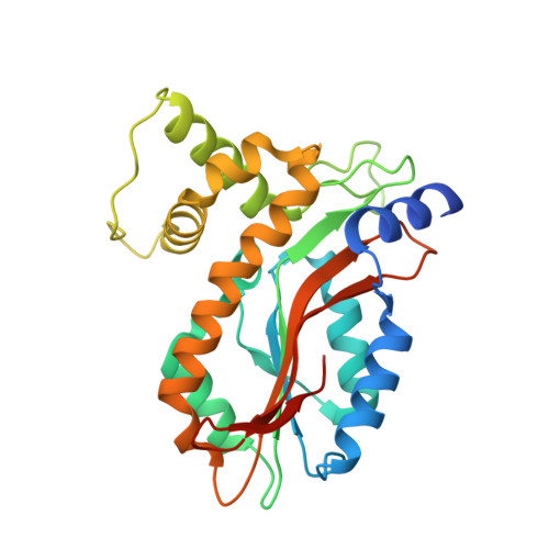
    6KFS

  • PubMed Abstract: 

    Carbonic anhydrases (CAs) are a well characterized family of metalloenzymes that are highly efficient in facilitating the interconversion between carbon dioxide and bicarbonate. Recently, CA activity has been associated with the LCIB (limiting CO 2 -inducible protein B) protein family, which has been an interesting target in aquatic photosynthetic microorganisms. To gain further insight into the catalytic mechanism of this new group of CAs, the X-ray structure of a highly active LCIB homolog (PtLCIB3) from the diatom Phaeodactylum tricornutum was determined. The CA activities of PtLCIB3, its paralog PtLCIB4 and a variety of their mutants were also measured. It was discovered that PtLCIB3 has a classic β-CA fold and its overall structure is highly similar to that of its homolog PtLCIB4. Subtle structural alterations between PtLCIB3 and PtLCIB4 indicate that an alternative proton-shuttle cavity could perhaps be one reason for their remarkable difference in CA activity. A potential alternative proton-shuttle route in the LCIB protein family is suggested based on these results.


  • Organizational Affiliation

    School of Biological Science, Nanyang Technological University, 60 Nanyang Drive, Singapore 637551, Singapore.


Macromolecules
Find similar proteins by:  (by identity cutoff)  |  3D Structure
Entity ID: 1
MoleculeChains Sequence LengthOrganismDetailsImage
LCIB_C_CA domain-containing protein267Phaeodactylum tricornutumMutation(s): 0 
Gene Names: pt43233
UniProt
Find proteins for B7FR28 (Phaeodactylum tricornutum (strain CCAP 1055/1))
Explore B7FR28 
Go to UniProtKB:  B7FR28
Entity Groups  
Sequence Clusters30% Identity50% Identity70% Identity90% Identity95% Identity100% Identity
UniProt GroupB7FR28
Sequence Annotations
Expand
  • Reference Sequence
Small Molecules
Ligands 1 Unique
IDChains Name / Formula / InChI Key2D Diagram3D Interactions
ZN (Subject of Investigation/LOI)
Query on ZN

Download Ideal Coordinates CCD File 
B [auth A]ZINC ION
Zn
PTFCDOFLOPIGGS-UHFFFAOYSA-N
Experimental Data & Validation

Experimental Data

  • Method: X-RAY DIFFRACTION
  • Resolution: 1.20 Å
  • R-Value Free: 0.167 
  • R-Value Work: 0.149 
  • Space Group: H 3 2
Unit Cell:
Length ( Å )Angle ( ˚ )
a = 93.363α = 90
b = 93.363β = 90
c = 177.834γ = 120
Software Package:
Software NamePurpose
PHENIXrefinement
XDSdata reduction
XDSdata scaling
PHASERphasing

Structure Validation

View Full Validation Report


Entry History & Funding Information

Deposition Data


Funding OrganizationLocationGrant Number
Ministry of Education (Singapore)SingaporeMOE2015-T2-1-078

Revision History  (Full details and data files)

  • Version 1.0: 2020-07-15
    Type: Initial release
  • Version 1.1: 2020-07-22
    Changes: Database references
  • Version 1.2: 2024-03-27
    Changes: Data collection, Database references