


Crystal Structure of DphMB1
Fan, S.To be published.
Experimental Data Snapshot
wwPDB Validation 3D Report Full Report

Entity ID: 1 | |||||
|---|---|---|---|---|---|
| Molecule | Chains | Sequence Length | Organism | Details | Image |
| Lipase | A [auth B], B [auth A], C, D | 320 | Mycobacterium adipatum | Mutation(s): 0 Gene Names: A7U43_17735, dphMB1 | 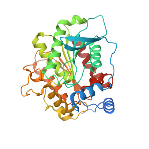 |
UniProt | |||||
Find proteins for A0A172UPQ1 (Mycobacterium adipatum) Explore A0A172UPQ1 Go to UniProtKB: A0A172UPQ1 | |||||
Entity Groups | |||||
| Sequence Clusters | 30% Identity50% Identity70% Identity90% Identity95% Identity100% Identity | ||||
| UniProt Group | A0A172UPQ1 | ||||
Sequence AnnotationsExpand | |||||
| |||||
| Length ( Å ) | Angle ( ˚ ) |
|---|---|
| a = 64.575 | α = 90 |
| b = 159.539 | β = 105.09 |
| c = 71.132 | γ = 90 |
| Software Name | Purpose |
|---|---|
| PHENIX | refinement |
| HKL-2000 | data reduction |
| Funding Organization | Location | Grant Number |
|---|---|---|
| National Natural Science Foundation of China | China | 31170119 |
RCSB PDB (citation) is hosted by
RCSB PDB is a member of the

