6K27

Pyrophosphatase with PPi from Acinetobacter baumannii


Experimental Data Snapshot

  • Method: X-RAY DIFFRACTION
  • Resolution: 1.86 Å
  • R-Value Free: 0.245 
  • R-Value Work: 0.196 
  • R-Value Observed: 0.196 

wwPDB Validation   3D Report Full Report


Ligand Structure Quality Assessment 


This is version 1.1 of the entry. See complete history


Literature

Crystal Structures of Pyrophosphatase from Acinetobacter baumannii: Snapshots of Pyrophosphate Binding and Identification of a Phosphorylated Enzyme Intermediate.

Si, Y.Wang, X.Yang, G.Yang, T.Li, Y.Ayala, G.J.Li, X.Wang, H.Su, J.

(2019) Int J Mol Sci 20

  • DOI: https://doi.org/10.3390/ijms20184394
  • Primary Citation of Related Structures:  
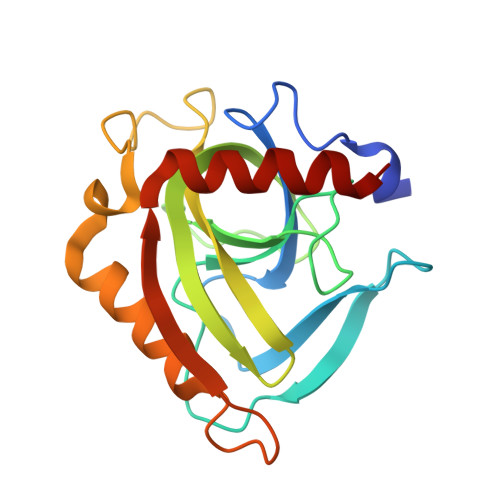
    6K21, 6K27, 6KI7, 6KI8

  • PubMed Abstract: 

    All living things have pyrophosphatases that hydrolyze pyrophosphate and release energy. This energetically favorable reaction drives many energetically unfavorable reactions. An accepted catalytic model of pyrophosphatase shows that a water molecule activated by two divalent cations (M1 and M2) within the catalytic center can attack pyrophosphate in an S N 2 mechanism and thus hydrolyze the molecule. However, our co-crystal structure of Acinetobacter baumannii pyrophosphatase with pyrophosphate shows that a water molecule from the solvent may, in fact, be the actual catalytic water. In the co-crystal structure of the wild-type pyrophosphatase with pyrophosphate, the electron density of the catalytic centers of each monomer are different from one another. This indicates that pyrophosphates in the catalytic center are dynamic. Our mass spectroscopy results have identified a highly conserved lysine residue (Lys30) in the catalytic center that is phosphorylated, indicating that the enzyme could form a phosphoryl enzyme intermediate during hydrolysis. Mutation of Lys30 to Arg abolished the activity of the enzyme. In the structure of the apo wild type enzyme, we observed that a Na + ion is coordinated by residues within a loop proximal to the catalytic center. Therefore, we mutated three key residues within the loop (K143R, P147G, and K149R) and determined Na + and K + -induced inhibition on their activities. Compared to the wild type enzyme, P147G is most sensitive to these cations, whereas K143R was inactive and K149R showed no change in activity. These data indicate that monovalent cations could play a role in down-regulating pyrophosphatase activity in vivo. Overall, our results reveal new aspects of pyrophosphatase catalysis and could assist in the design of specific inhibitors of Acinetobacter baumannii growth.


  • Organizational Affiliation

    Jilin Province Key Laboratory on Chemistry and Biology of Natural Drugs in Changbai Mountain, School of Life Sciences, Northeast Normal University, Changchun 130024, China.


Macromolecules
Find similar proteins by:  (by identity cutoff)  |  3D Structure
Entity ID: 1
MoleculeChains Sequence LengthOrganismDetailsImage
Inorganic pyrophosphatase
A, B, C, D, E
A, B, C, D, E, F, G, H
177Acinetobacter baumanniiMutation(s): 1 
Gene Names: ppa
EC: 3.6.1.1
UniProt
Find proteins for A0A0A7XPH7 (Acinetobacter nosocomialis)
Explore A0A0A7XPH7 
Go to UniProtKB:  A0A0A7XPH7
Entity Groups  
Sequence Clusters30% Identity50% Identity70% Identity90% Identity95% Identity100% Identity
UniProt GroupA0A0A7XPH7
Sequence Annotations
Expand
  • Reference Sequence
Small Molecules
Ligands 2 Unique
IDChains Name / Formula / InChI Key2D Diagram3D Interactions
DPO (Subject of Investigation/LOI)
Query on DPO

Download Ideal Coordinates CCD File 
CA [auth F]
GA [auth G]
I [auth A]
KA [auth H]
M [auth B]
CA [auth F],
GA [auth G],
I [auth A],
KA [auth H],
M [auth B],
Q [auth C],
U [auth D],
Y [auth E]
DIPHOSPHATE
O7 P2
XPPKVPWEQAFLFU-UHFFFAOYSA-J
MG (Subject of Investigation/LOI)
Query on MG

Download Ideal Coordinates CCD File 
AA [auth E]
BA [auth E]
DA [auth F]
EA [auth F]
FA [auth F]
AA [auth E],
BA [auth E],
DA [auth F],
EA [auth F],
FA [auth F],
HA [auth G],
IA [auth G],
J [auth A],
JA [auth G],
K [auth A],
L [auth A],
LA [auth H],
MA [auth H],
N [auth B],
NA [auth H],
O [auth B],
P [auth B],
R [auth C],
S [auth C],
T [auth C],
V [auth D],
W [auth D],
X [auth D],
Z [auth E]
MAGNESIUM ION
Mg
JLVVSXFLKOJNIY-UHFFFAOYSA-N
Experimental Data & Validation

Experimental Data

  • Method: X-RAY DIFFRACTION
  • Resolution: 1.86 Å
  • R-Value Free: 0.245 
  • R-Value Work: 0.196 
  • R-Value Observed: 0.196 
  • Space Group: H 3
Unit Cell:
Length ( Å )Angle ( ˚ )
a = 110.294α = 90
b = 110.294β = 90
c = 302.492γ = 120
Software Package:
Software NamePurpose
PHENIXrefinement

Structure Validation

View Full Validation Report


Ligand Structure Quality Assessment 


Entry History 

Deposition Data

  • Released Date: 2019-10-02 
  • Deposition Author(s): Su, J.

Revision History  (Full details and data files)

  • Version 1.0: 2019-10-02
    Type: Initial release
  • Version 1.1: 2024-03-27
    Changes: Data collection, Database references, Derived calculations